Figures & data
Figure 1. Sagittal (A) and axial (B) MRI after intravenous application of gadolinium. The L5 tumor protruded into the epidural space where it compressed the right L5 nerve root.
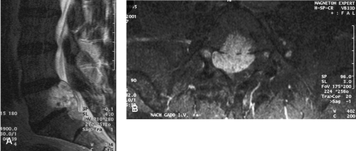
Figure 2. Sagittal (A) and axial (B) T2-weighted MRI images showing tumor recurrence (arrow) after posterior resection.
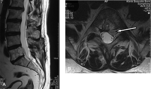
Figure 3. Anteroposterior (A) and lateral (B) radiographs showing circular reconstruction after L5 total spondylectomy.
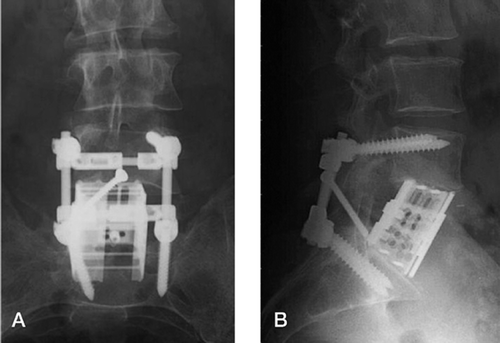
Figure 4. A. sagittal CT reconstruction depicting solid bony fusion through the cage and physiological alignment. B. Axial CT showing the cage filled with bone (horizontal arrow) and the posterior implants (vertical arrow) 6 months after surgery.
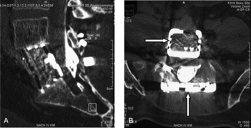
Figure 5. Axial MRI after application of contrast medium showing the second recurrence of the tumor (between arrows).
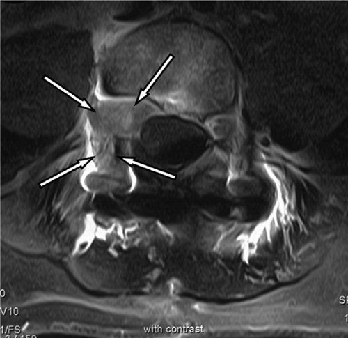
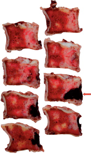
Figure 6. Multiple macroscopic sections of the L5 VB. The pedicles were removed during the first stage of the operation. The arrow indicates the epicentre of the chordoma in the posterior part of the VB where it crossed the posterior wall.