Figures & data
Figure 1. Photomicrograph of fibroblast colony-forming units obtained from cultured iliac bone marrow mononuclear cells (Wright's stain, magnification × 160).
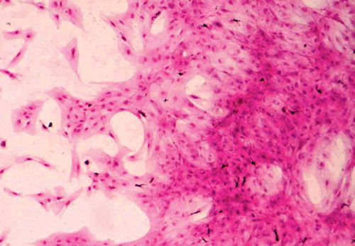
Figure 2. Graph showing a comparison of the mean number of fibroblast colony-forming units (CFU-F) per 4 × 10⁵ bone marrow mononuclear cells in iliac and tibial marrow from each patient. Each point shows the mean number of CFU-F for triplicate assays. Data from individual patients are connected by broken lines.
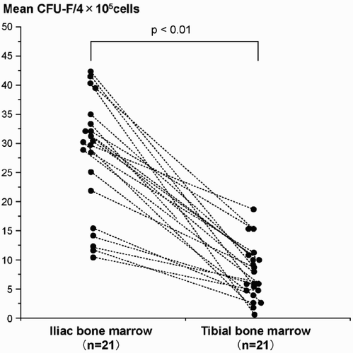
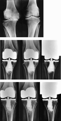
Figure 3. A. Preoperative radiograph of the knees of a patient with osteoarthritis who underwent simultaneous bilateral total knee arthroplasty. B. Serial postoperative radiographs of the knee that received total knee arthroplasty with autologous bone marrow grafting. Note the presence of an initial gap at the tibial tray-bone interface at 2 weeks after surgery (left panel). However, the gap is no longer seen 6 months (middle panel) and 1 year after surgery (right panel). C. Serial postoperative radiographs of the contralat C.Serial postoperative radiographs of the contralateral knee, which received total knee arthroplasty without autologous bone marrow grafting. Note the presence of an initial gap at the tibial tray-bone interface at 2 weeks after surgery (left panel). This gap persisted 6 months (middle panel) and 1 year (right panel) after surgery.
Comparison of the number of prostheses with radiolucent lines and gaps in serial fluoroscopically-guided anteroposterior or lateral radiographs between the knees that underwent bone marrow grafting and those that did not