Figures & data
Table 1. Categorization of 26 piglets according to postoperative periods
Figure 1. Histological specimen of right (Rt) and left (Lt) femurs extracted from a piglet killed at postoperative week 4.The left femoral head (Lt) showed avascular necrosis of the capital femoral epiphysis.Marked reduction in size of ossific nucleus (*) and considerable hyperplasia of articular cartilage (arrow) were seen in the operated femur. (Hematoxylin and eosin, ×1).
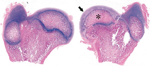
Figure 2. Gross (A) and radiographic (B) findings from extracted femurs from a piglet killed at postoperative week 20 showing (to the right) decreased epiphyseal height (44%), generalized flattening of the articular surface, coxa magna (16%), and severe GT overgrowth.
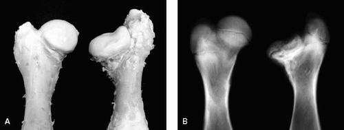
Table 2. Responses of the femoral head in 26 piglets with vascular infarct
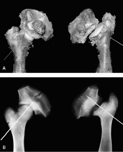
Figure 3. Gross (A) and radiographic (B) findings from extracted femurs from a piglet sacrificed at postoperative week 20 showing (to the right) subluxation of the femoral head with severe collapse of head and dysplasia of the acetabulum.