Figures & data
Figure 1 a. Preoperative lateral view. b. Preoperative anteroposterior view. Pre- and postoperative radiographs of the GSB III elbow arthroplasty in a 58-year-old woman with severe rheumatoid destruction of the elbow joint.
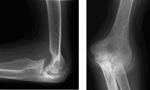
Figure 2 a. Postoperative lateral view. The arrows mark adequate cement mantle around the ulnar stem extending past the tip, and the inadequate cement mantle of the humeral stem, which does not extend past the tip of the component. b. Postoperative anteroposterior view. Note the preservation of the distal humeral condyles with muscular and ligamentous attachments, thereby improving soft tissue stability of the arthroplasty and reduction of peak forces at the interfaces. The radial head has routinely been removed. Pre- and postoperative radiographs of the GSB III elbow arthroplasty in a 58-year-old woman with severe rheumatoid destruction of the elbow joint.
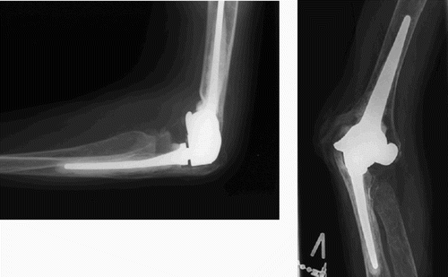
Table 1. Range of movement (ROM), preoperatively and postoperatively
Table 2. Results of the cementing technique
Table 3. Radiological signs of aseptic loosening at the latest follow-up