1,234
Views
13
CrossRef citations to date
0
Altmetric
Original papers

Modified pubic osteotomy for medialization of the femoral head in periacetabular osteotomy: A retrospective study of 144 hips

, , , &
Pages 474-482 | Received 05 Apr 2007, Accepted 10 Jan 2008, Published online: 08 Jul 2009

Figures & data

Figure 1. Cutting angle of pubic osteotomy (black line). The angle of the pubic osteotomy (right hip) is modified, with a 30‐degree inclination to the horizontal line (M group). The conventional pubic osteotomy (left hip) has a 90‐degree inclination to the horizontal line (C group).
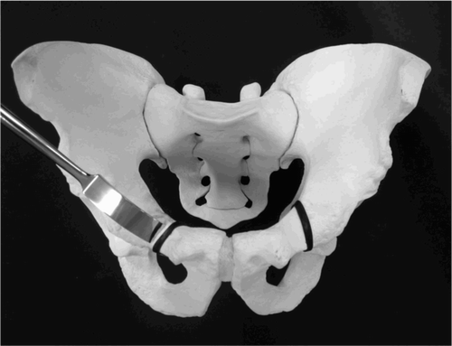
Figure 1. Cutting angle of pubic osteotomy (black line). The angle of the pubic osteotomy (right hip) is modified, with a 30‐degree inclination to the horizontal line (M group). The conventional pubic osteotomy (left hip) has a 90‐degree inclination to the horizontal line (C group).

Figure 2. Pelvic radiographic parameters: interhip distance (l), pelvic width (C), pelvic height (H), radius of the femoral head (r), center‐edge angle of Wiberg (θCE), and the frontal coordinates of point T (the midpoint of the straight line connecting the most lateral point (point 1) with the highest point (point 2) on the greater trochanter) were used as input data for computation of the resultant hip force R.
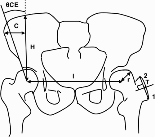
Figure 2. Pelvic radiographic parameters: interhip distance (l), pelvic width (C), pelvic height (H), radius of the femoral head (r), center‐edge angle of Wiberg (θCE), and the frontal coordinates of point T (the midpoint of the straight line connecting the most lateral point (point 1) with the highest point (point 2) on the greater trochanter) were used as input data for computation of the resultant hip force R.

Figure 3. Radiographic indices measured from AP radiographs: Kohler's ilioischial line (b‐c), distance between the bilateral Kohler ilioischial lines (t), and distance from each Kohler ilioischial line to the center of the femoral head (a). Head laterailzation index (HLI) was calculated using the following formula: HLI = a /t / 2.
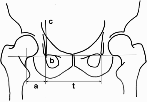
Figure 3. Radiographic indices measured from AP radiographs: Kohler's ilioischial line (b‐c), distance between the bilateral Kohler ilioischial lines (t), and distance from each Kohler ilioischial line to the center of the femoral head (a). Head laterailzation index (HLI) was calculated using the following formula: HLI = a /t / 2.

Figure 4. Medialization type. (A) A 24‐year‐old female with Tönnis classification grade 1 OA before CPO in 2006. The CE angle and ARO are 5° and 22°, respectively. (B) Immediately after surgery, showing a modified pubic osteotomy with a 30° inclination from the horizontal line in CPO. (C) Bony union at the osteotomy sites is observed at 4 months after CPO. The CE angle and ARO are 44° and ‐7°, respectively. Since the ratio of lateralization of the femoral head is 0.73, this hip is classified as the medialization type.
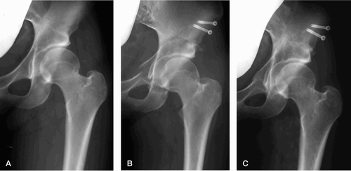
Figure 4. Medialization type. (A) A 24‐year‐old female with Tönnis classification grade 1 OA before CPO in 2006. The CE angle and ARO are 5° and 22°, respectively. (B) Immediately after surgery, showing a modified pubic osteotomy with a 30° inclination from the horizontal line in CPO. (C) Bony union at the osteotomy sites is observed at 4 months after CPO. The CE angle and ARO are 44° and ‐7°, respectively. Since the ratio of lateralization of the femoral head is 0.73, this hip is classified as the medialization type.

Figure 5. Lateralization type. (A) An 18‐year‐old female with Tönnis classification grade 1 OA before CPO in 2004. The CE angle and ARO are 7° and 14°, respectively. (B) Immediately after surgery, showing a conventional pubic osteotomy performed vertically to the horizontal line in CPO. (C) The CE angle and ARO have improved to 30° and ‐2°, respectively. Since the ratio of lateralization of the femoral head is 1.16, this hip is classified as the lateralization type.
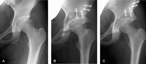
Figure 5. Lateralization type. (A) An 18‐year‐old female with Tönnis classification grade 1 OA before CPO in 2004. The CE angle and ARO are 7° and 14°, respectively. (B) Immediately after surgery, showing a conventional pubic osteotomy performed vertically to the horizontal line in CPO. (C) The CE angle and ARO have improved to 30° and ‐2°, respectively. Since the ratio of lateralization of the femoral head is 1.16, this hip is classified as the lateralization type.

Table 1.  Radiographic evaluations. Values are mean (SD)

Table 2.  Distribution of postoperative femoral head centers

Table 3.  Radiographic and mechanical parameters for the three displacement types. Values are mean (SD)

Table 4.  Radiographic and biomechanical parameters for the M and C groups. Values are mean (SD)

Reprints and Corporate Permissions

Please note: Selecting permissions does not provide access to the full text of the article, please see our help page How do I view content?

To request a reprint or corporate permissions for this article, please click on the relevant link below:

Academic Permissions

Please note: Selecting permissions does not provide access to the full text of the article, please see our help page How do I view content?

Obtain permissions instantly via Rightslink by clicking on the button below:

If you are unable to obtain permissions via Rightslink, please complete and submit this Permissions form. For more information, please visit our Permissions help page.