Figures & data
Figure 1. A. Sketch of the deformity and explanation of the nomenclature transferred from the lower limb to the forearm. IR: internal rotation; ER: external rotation; Pro: pronation; Sup: supination. B. Frame assembly with a proximal reference ring and the struts turned upside down for better visualization of the patient. The Master Tab is 0° anterior. Transfixation of the wrist is removed after lengthening.
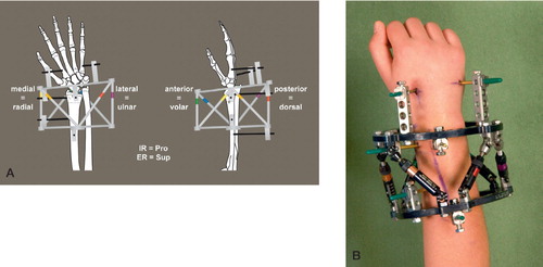
Figure 2. Patient 1. A and B. Pseudo‐Madelung deformity of the right distal radius after physial injury and closed radial growth plate. At the time of correction, the ulnar growth plate was in stage G according to the Tanner and Whitehouse scale. C and D. After deformity correction, with an almost normal functioning wrist and anatomically reduced DRUJ 1 year after correction.
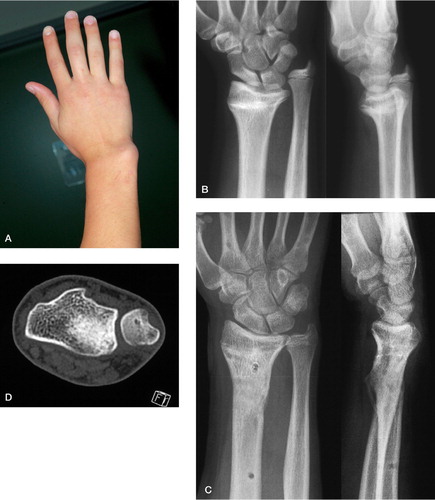
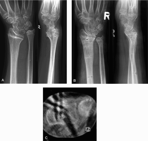
Figure 3. Patient 2. Before (A) and after (B) correction of the distal radius deformity of the right forearm. The radial growth plate was partially closed (> 50%) at the time of correction and the ulnar growth plate was at stage G according to the Tanner and Whitehouse scale. The DRUJ was reduced anatomically in the low‐dose CT at the end of correction (C).
Radial deformities, distraction data and ROM