Figures & data
Figure 1. Illustrations of the meniscal root tear classification system in 5 different groups based on tear morphology. For consistency, all meniscal tears are shown as medial meniscal posterior root tears in this illustration. The classification of 5 tear patterns was based on morphology: partial stable root tear (type 1), complete radial tear within 9 mm of the bony root attachment (type 2), bucket-handle tear with complete root detachment (type 3), complex oblique or longitudinal tear with complete root detachment (type 4), and bony avulsion fracture of the root attachment (type 5). Reprinted with permission from LaPrade et al. (Citation2015b).
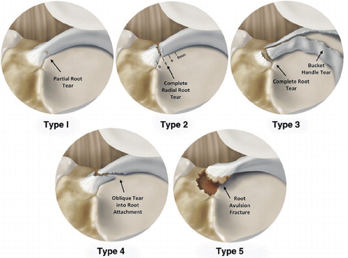
Table 1. Classification of anterior and posterior tears of the medial and lateral meniscal roots based on tear morphology
Figure 2. Cadaveric image (superior view) demonstrating the anatomical landmarks to identify a medial meniscus posterior root attachment (asterisk) in a right knee. MTE: medial tibial eminence; MARA: medial meniscus anterior root attachment; LARA: lateral meniscus anterior root attachment; MPRA: medial meniscus posterior root attachment; LPRA: lateral meniscus posterior root attachment.
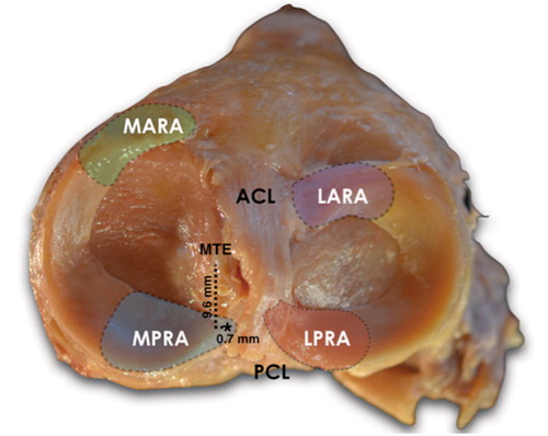
Figure 3. Right knee image demonstrating the close relationship between the posterior root attachments and the PCL.
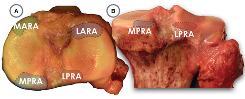
Figure 4. Pertinent anatomical relationships (right knee) as reported by Johannsen et al.7 A. Superior view. B. Posterior view. Reprinted with permission from Johannsen et al. (Citation2012). ACL: anterior cruciate ligament bundle attachments; LPRA: lateral meniscus posterior root attachment; LTE: lateral tibial eminence; MPRA: medial meniscus posterior root attachment; MTE: medial tibial eminence; PCL: posterior cruciate ligament bundle attachments; SWF: shiny white fibers of posterior horn of medial meniscus.
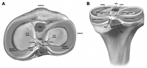
Figure 5. Visualization of meniscal root tears via magnetic resonance imaging. A. Coronal T2-weighted section demonstrating medial meniscal extrusion (arrow) (left knee). B. Axial image demonstrating high signal in region of meniscus root and posterior horn with a radial root tear (arrow) (right knee). C. Sagittal image demonstrating ghost sign (arrow) (right knee). Reprinted with permission from Bhatia et al. (Citation2014).
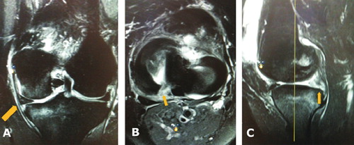
Figure 6. Steps taken during arthroscopic repair of the posterior medial meniscus radial root (left knee). A. Radial root tear. B. Probing of root tear through posteromedial knee portal cannula. C. Placement of shuttle suture device through body of root tear. D. Transosseous pullout repair of root tear. E. Pullout sutures tied over a button on the anteromedial tibia. Reprinted with permission from Bhatia et al. (Citation2014).
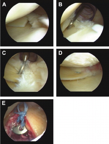
Figure 7. Preferred technique for fixation of a posterior horn medial meniscal root tear involves transosseous suture repair tied over a button on the anteromedial tibia. Proper tensioning and anatomical placement of the attachment are critical for healing and restoration of meniscal function. Reprinted with permission from Padalecki et al. (Citation2014).
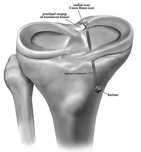
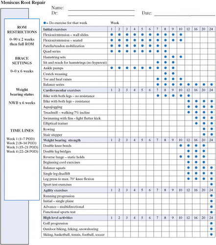
Figure 8. The authors’ rehabilitation protocol after posterior meniscal root repair.