
Figures & data
Figure 1. A 9-year-old boy sustained a radial neck fracture (Judet type-IV) with remaining bony contact of the radial neck, after falling on a level surface (panels a and b). Panels c and d show radiographs 16 days after elastic stable intramedullary nailing (ESIN). After implant removal 3 months postoperatively, there was correct positioning of the radial head and neck (panels e and f). At clinical follow-up 2.3 years after the initial trauma, the patient was free of symptoms and had full range of motion.
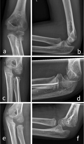
Figure 2. A 7-year-old girl had a type-B lesion after falling on a level surface (panels a and b). Note the hardly visible radial head on conventional radiographs (arrow). A CT scan (panels c and d) revealed the location of the radial head. Open reduction and elastic stable intramedullary nailing (ESIN) was necessary. 19 months later, there were signs of shortening and angulation but no evidence of avascular necrosis (panels e and f). Clinically, the patient was free of symptoms, with full range of motion and a Linscheid-Wheeler score of I.
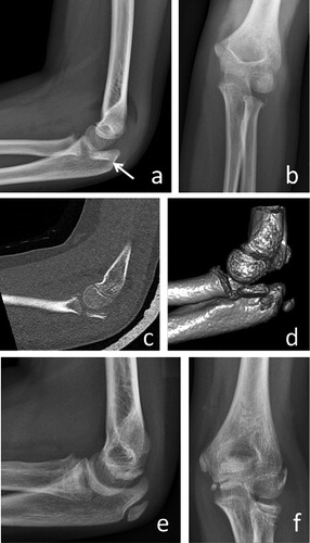
Table 1. Comparison of age, gender distribution, and additional injuries in group A (bony contact) and group B (no bony contact)
Table 2. Demographic data, gender distribution, fracture type, treatment, Linscheid and Wheeler score, and length of follow-up
Table 3. Outcome in 19 children with radial neck fractures