
Figures & data
Figure 1. A. A 6-year-old boy with a Gartland grade-III extension-type supracondylar fracture. B and C. Satisfactory reduction and pin fixation in both the frontal plane (Baumann angle) (B) and the sagittal plane (C) (AHL crosses ossification center of capitellum).
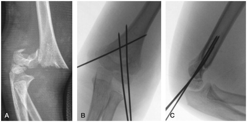
Figure 2. Pin-fixed supracondylar humerus fractures at the Children’s Hospital, Helsinki.
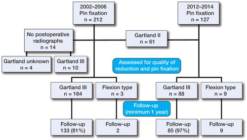
Table 1. The quality of reduction and pin fixation
Figure 3. The intersection point of the anterior humeral line with the ossification center of capitellum divided into 5 different zones.
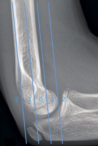
Table 2. The number of patients and number of postoperative radiographs per patient
Table 3. Timing of postoperative radiography
Figure 4. Quality of reduction of supracondylar humerus fracture in 264 children. Green: satisfactory alignment; yellow and red: unsatisfactory alignment; red: 10˚ outside normal values.
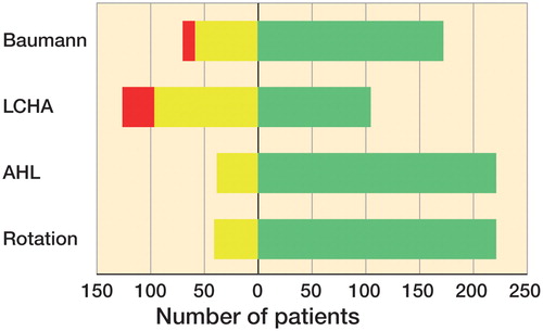
Table 4. The intersection point of the anterior humeral line with the ossification center of the capitellum in 5 different zones
Figure 5. A. AP elbow radiograph of a 6-year-old boy with Gartland grade-III extension-type supracondylar fracture. B. Malunion at 3 weeks after unsatisfactory reduction and pin fixation in AP and sagittal planes. C. Corrective osteotomy was scheduled 3 years later after 7 postoperative radiographs.
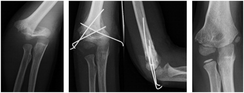
Table 5. The quality of reduction and pin fixation in follow-up patients and non-follow-up patients during the 2 study periods (2002–2006 and 2012–2014)