
Figures & data
Figure 1. At 6 weeks. MRI (A) and contrast MRI (B). Prior to stage Ia.
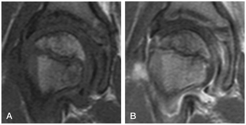
Figure 2. At 2.3 months. Start of extrusion. Subchondral fracture. Early stage Ia.
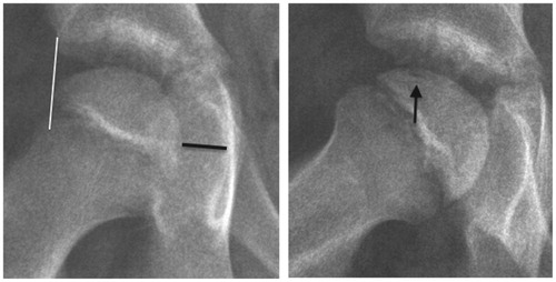
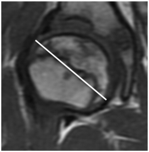
Figure 3. At 5.2 months. Contrast MRI. Cyst. Stage Ib. 2 days later, denosumab was administered.
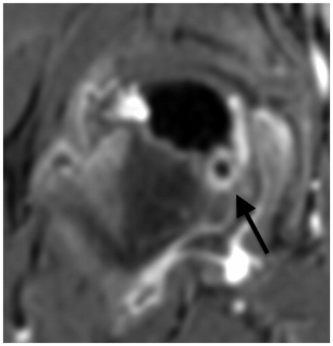
Figure 4. At 11.5 months. PD-weighted MRI. Beginning of loss of containment. Stage IIb.
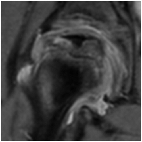
Figure 5. At 1 year. Moderate extrusion. Anterior flattening. Stage IIIa.
A proximal varus osteotomy was performed 1 month later.
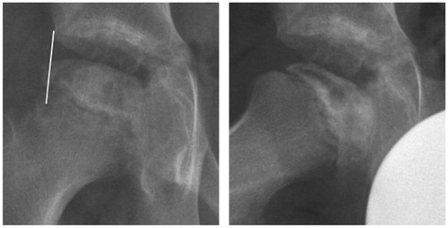
Figure 6. Six weeks postoperatively.
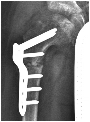
Figure 7. At 2 years. 90% re-ossification. Stage IIIb.
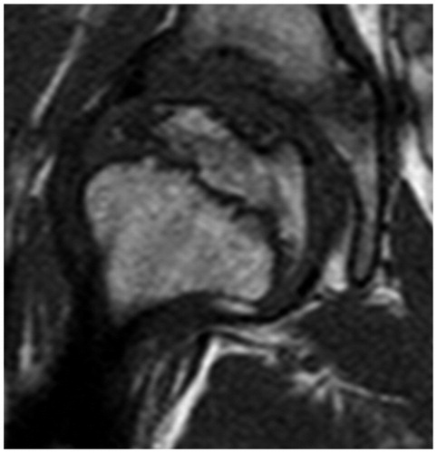
Figure 8C. At 2 years and 8 months. An 11% increase in the maximum diameter. Almost stage IV.
Supplemental material