Figures & data
Figure 1 a. Preoperative picture of the patient at the age of 13 years, showing pronounced varus deformity of her right leg.
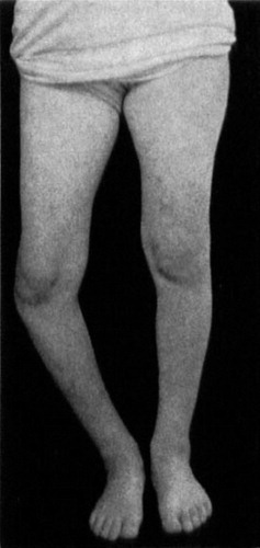
Figure 1 b. Preoperative radiographs, without weight-bearing (left). During weight-bearing (right) varus is increased because the defective medial condyle provides poor support for the femoral condyle. Reprinted from Støren (1969).
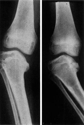
Figure 1 c. Operative technique. The chiseling is done proximal to the epiphyseal line and proceeds to the midline (left). The fragment is slowly elevated, lifting the whole fragment in one piece (middle). Solid bone grafts from the iliac crest, large enough to force the fragment into maximal elevation, are wedged under the fragment under maximal forced valgus of the knee (right).
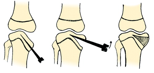
Figure 1 d. Radiograph 1 year after operative elevation of the medial tibial joint surface, showing consolidation and good correction of the varus deformity.
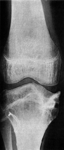
Figure 2 a. Pictures at 65 years of follow-up (patient age 78 years), showing slight varus deformity of the right leg.
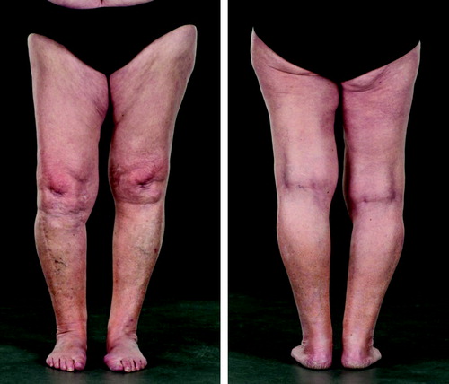
Figure 2 b. At 65 years follow-up the patient had almost full knee flexion.
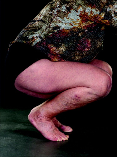
Figure 2 c. Radiographs at 65 years follow-up, showing moderate osteoarthritis of the right knee.
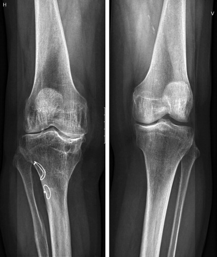
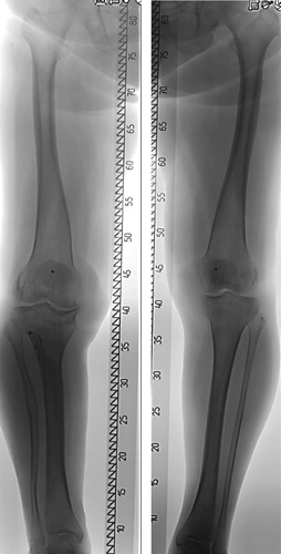
Figure 2 d. Long-leg standing radiographs at 65 years follow-up, showing slight varus of the right knee and leg length discrepancy of 1.5 cm with the right leg shorter.