
Figures & data
Figure 1. Coronal (A) and axial (B) BFFE MR images (0.5 mm) in a 3-month-old boy (patient 18) with brachial plexus birth injury on the right side. Total (both ventral and dorsal roots) avulsion of right C6 root with a PMC (asterisk): ventral root is avulsed from the cord (upper arrow), where a short stump of dorsal root is seen (lower arrow). Left ventral and dorsal C6 roots (arrowheads) are normal.
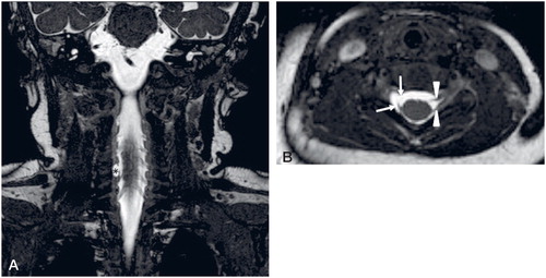
Figure 2. Axial BFFE MR image (0.5 mm) in a 4-month-old girl (patient 26) with brachial plexus birth injury on the right side. Partial avulsion of C8 root: ventral root is avulsed (red arrow), dorsal C8 root is intact (arrowhead). Left C8 nerve roots are normal (arrowheads).
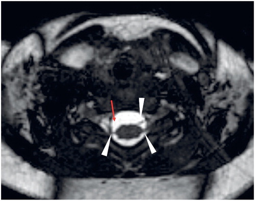
Figure 3. Coronal BFFE image (0.5 mm) in a 3-month-old girl (patient 16) with brachial plexus birth injury on the right side. Right ventral C6 root is thinned (arrow) compared to the normal left ventral C6 root (arrowhead).
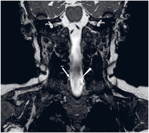
Figure 4. Coronal (A) and axial (B) BFFE image (0.5 mm) in 3-month-old boy (patient 31) with brachial plexus birth injury on both sides. (b) Intact ventral and dorsal nerve roots (red arrows) at C7 level despite a PMC clearly visible on both sides.
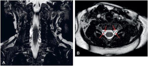
Table 1. Patient demographics. Patients are arranged in descending order of abnormal findings on MRI
Figure 5. Multivariable model expressing GSA difference in relation to age at time of MRI and clinical findings at birth.
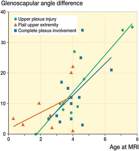
Table 2. Glenohumeral joint (GHJ) MRI findings. Patients are arranged primarily in descending order based on incongruency of their affected shoulder, secondarily in descending order based on the difference between GSA of both shoulders
Table 3. MRI findings compared to intra-operative findings by root level
Table 4. Patient outcome. Outcome expressed as ratio (%) of active antigravity range of motion of the affected side in comparison with the unaffected side. Patients are arranged primarily by the extent of injury at birth and secondarily by their 3-month Test Score