Figures & data
Figure 1. LDFA and MPTA comparing preoperative and postoperative distributions.
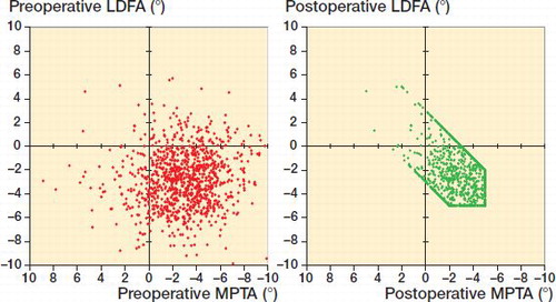
Table 1. Lower limb alignment of pre-operative anatomy compared with after rKA. Values are mean (SD) [range] degrees.
Figure 2. Distribution of medio-lateral gap imbalance in the extension space for rKA and MA techniques (p < 0.001).
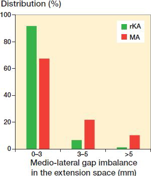
Table 2. Distribution of medial and lateral gap sizes in the extension space for MA and rKA techniques. Values are percentages
Table 3. Medial and lateral gaps modification in the extension space and resulting medio-lateral difference in mm for MA and rKA techniques. Values are mean (SD) [range]
Figure 3. Distribution of medio-lateral gap imbalance in the flexion space for rKA and MA with PC 3° (p < 0.001) or TEA (p < 0.001) techniques.
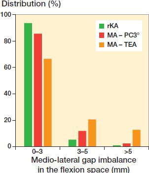
Table 4. Medial and lateral gaps modification in the flexion space and resulting medio-lateral difference in mm for MA PC method, MA TEA method, and rKA techniques. Values are mean (SD) [range]
Table 5. Flexion–extension gap differences (ΔFE) in mm for the medial and lateral compartments for MA PC method, MA TEA method, and rKA techniques. Values are mean (SD) [range]
Table 6. Percentage of knees with medial or lateral flexion-extension gap mismatch for MA PC method, MA TEA method, and rKA techniques
Table 7. Percentage of knees where the medio-lateral gap mismatch is present in both the extension and flexion spaces for MA PC method, MA TEA method and rKA techniques
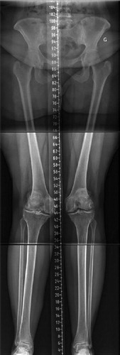
Figure 4. Lower limb long radiographs showing a case with an LDFA of 11° and MPTA of 6°. Reproducing her lower limb alignment with KA technique (unrestricted) would leave her lower limb HKA in 5° of valgus. With rKA, correcting the femur to 5° and the tibia to 2° of varus would results in an HKA of 3° valgus.