
Figures & data
Figure 1. Compress components with distal femoral adaptor.
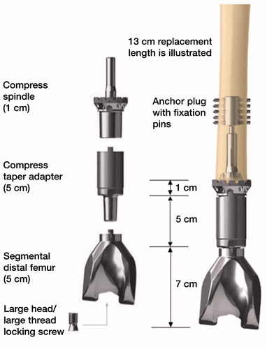
Figure 2. Training and test cohort protocol.
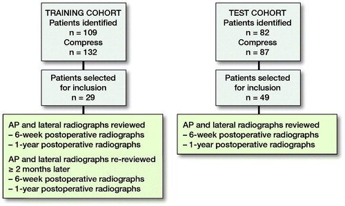
Figure 3. Radiographic parameters used to construct the fast and frugal tree model.
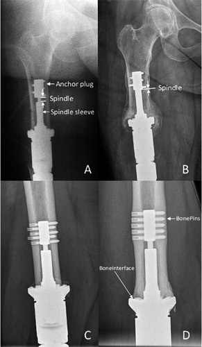
Figure 4. Training cohort FFT.

Figure 5. Test cohort FFT.
A. Immediate postoperative radiograph of distal femoral reconstruction showing the initial length between the top of the spindle sleeve and bottom of anchor plug (Spindle).
B. 1-year postoperative radiograph of the same patient as in A, depicting the loss of length of the Spindle.
C. Immediate postoperative radiograph of distal femoral reconstruction.
D. 1-year postoperative radiograph of the same patient as in , depicting hypertrophy at both the bone–implant interface (BoneInterface) and at the bone–pin interface (BonePins).
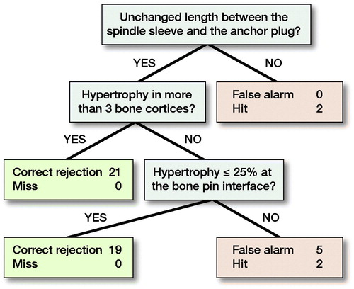
Table 1. Training cohort: test–retest intra-rater reliability
Table 2. Training cohort: inter-rater reliability