Figures & data
Figure 1. Type of Hohmann retractor used during the procedure.
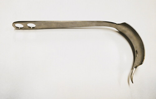
Figure 2. Hohmann retractors placed on a pelvis phantom.
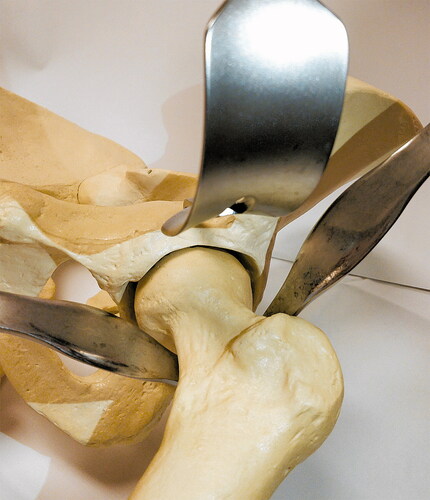
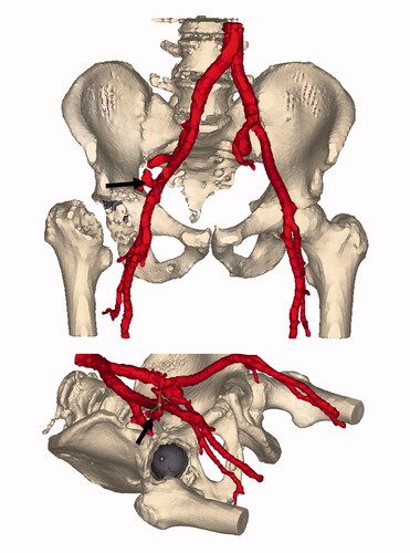
Figure 3. Segmentation of the contrast-enhanced vascular CT. Window ranges corresponding to intravascular contrast are colored red. The implanted metal backed cup is colored gray. The arrow is placed on the site of extravasation at the external iliac artery. Of note is the distance between the acetabular rim and the extravasation site.