
Figures & data
Figure 1. Diagram depicting the patient population and the reasons for inclusion in our cohort.
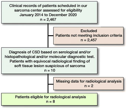
Figure 3. Case no. 7. A 40-year-old female with an epifascial soft tissue lesion of the upper arm. (A) On a T1-weighted image, the lesion with intermediate signal intensity and irregular margin; wide contact with the underlying fascia (arrow). (B) Homogenous high T2-weighted signal intensity without any surrounding edema (arrow). (C) Coronal T1-weighted image with fat saturation after application of Gd-contrast shows heterogeneous contrast enhancement (arrow). (D–E) DWI image (D) with corresponding (E) ADC map shows diffusion restriction due to necrotic collection (circles).
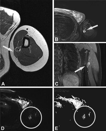
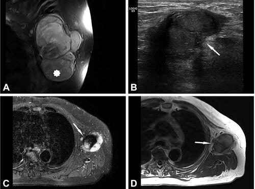
Table 1. Summary of clinical, serological, pathological and molecular data
Table 2. MRI characteristics of the lesions with differential diagnosis
Table 3. Summary of treatment and follow-up