3,755
Views
1
CrossRef citations to date
0
Altmetric
Original Article

On the diagnosis and treatment of femoral neck fractures

, MD
Pages 1-26 | Published online: 18 Nov 2009

Figures & data

Figure 1. The hip joint and the proximal femur. Reprinted with permission from the BMJ.Citation[1]

Figure 1. The hip joint and the proximal femur. Reprinted with permission from the BMJ.Citation[1]

Text box 1. Signs and symptoms of a hip fracture

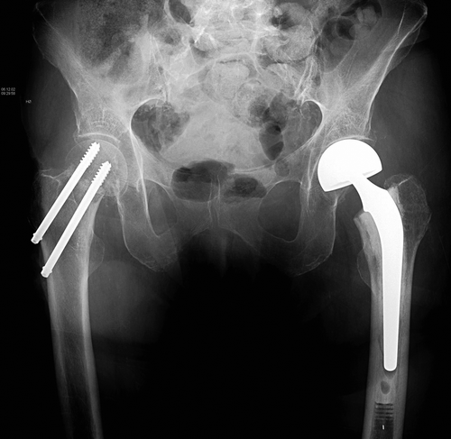
Text box 2. Different hip fractures and their treatment

Text box 3. Choices of internal fixation or arthroplastyCitation[9]

Figure 2. Garden's classification. Garden stages 1–4. Reproduced with permission and copyright © of the British Editorial Society of Bone and Joint Surgery Citation[80];Citation[85]

Figure 2. Garden's classification. Garden stages 1–4. Reproduced with permission and copyright © of the British Editorial Society of Bone and Joint Surgery Citation[80];Citation[85]

Figure 3. Anterioposterior pelvic radiograph of a patient who has suffered femoral neck fractures on both sides, only a few weeks apart. On the right side she was operated with two parallel screws and on the left side with a hemiarthroplasty.

Figure 3. Anterioposterior pelvic radiograph of a patient who has suffered femoral neck fractures on both sides, only a few weeks apart. On the right side she was operated with two parallel screws and on the left side with a hemiarthroplasty.

Figure 4. Example of screw voids in the femoral head after reversing the screws. The whiter appearance of the bone on the right hand side of the femoral head indicates that the bone density is higher in the femoral head near the joint space than it is towards the femoral neck. The better screw purchase provided by this bone is one of the reasons that the recommended screw placement is as near to the joint line as possible. The screws are only partially threaded to allow for compression over the fracture.

Figure 4. Example of screw voids in the femoral head after reversing the screws. The whiter appearance of the bone on the right hand side of the femoral head indicates that the bone density is higher in the femoral head near the joint space than it is towards the femoral neck. The better screw purchase provided by this bone is one of the reasons that the recommended screw placement is as near to the joint line as possible. The screws are only partially threaded to allow for compression over the fracture.

Reprints and Corporate Permissions

Please note: Selecting permissions does not provide access to the full text of the article, please see our help page How do I view content?

To request a reprint or corporate permissions for this article, please click on the relevant link below:

Academic Permissions

Please note: Selecting permissions does not provide access to the full text of the article, please see our help page How do I view content?

Obtain permissions instantly via Rightslink by clicking on the button below:

If you are unable to obtain permissions via Rightslink, please complete and submit this Permissions form. For more information, please visit our Permissions help page.