Figures & data
Table 1. Composition of gels.
Figure 1. DSC thermograms of Tenoxicam and final gel formulation.
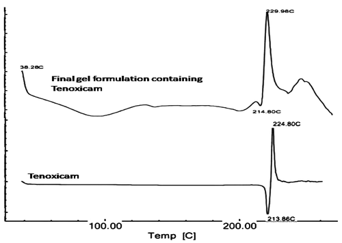
Figure 2. Transmission electron microscope photographs of (T3) Tenoxicam-loaded ultradeformable vesicles (magnification × 100,000).
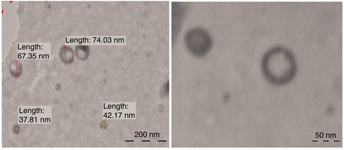
Figure 3. Effect of concentrations of different surfactants on the size of ultradeformable vesicles. Notes: S.D.C. is sodium desoxycholate. *Significant (p < 0.05) compared to T3.
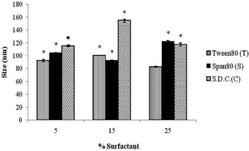
Figure 4. Effect of concentrations of different surfactants on the deformability of ultradeformable vesicles. Note: *Significant (p < 0.05) compared to T3.
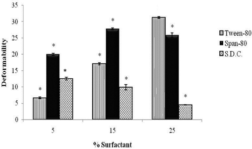
Table 2. Comparative kinetic analysis of vesicles: release rate constants (k), correlation coefficients (R 2), and release exponent (n) of different ultradeformable vesicles.
Table 3. Comparative features of different ultradeformable vesicles (n = 3).
Figure 5. Comparative release profile of optimised ultradeformable vesicle suspension and free drug solution.
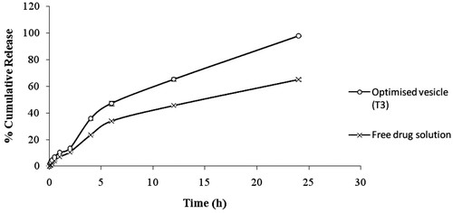
Figure 6. Frequency sweep curves of different gel formulations.
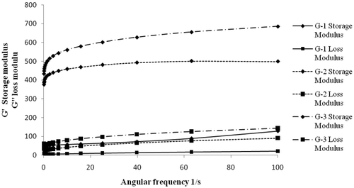
Table 4. Comparative kinetic analysis of gels: release rate constants (k), correlation coefficients (R 2), and release exponent (n) of gels of different concentrations.
Figure 7. Comparative release profile (dialysis membrane) of optimised gel formulation and free drug gel (n = 3).
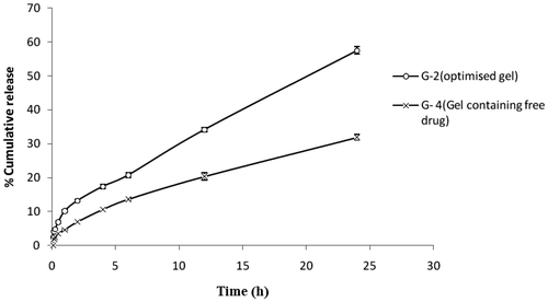
Figure 8. Comparative release profile (rat skin) of optimised gel formulation and free drug gel (n = 3).
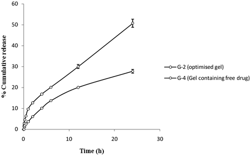
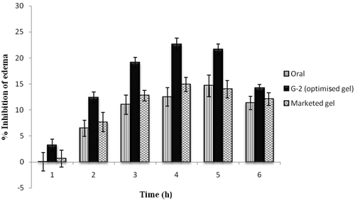
Figure 9. Comparative pharmacodynamic analysis of Tenoxicam ultradeformable vesicle G-2, Tenoxicam oral suspension in distilled water and marketed aceclofenac gel (Hifenac®) formulation by the carrageenan-induced rat paw oedema model.