Figures & data
Figure 1. XRD patterns of the sample CoFe1.9Ho0.1O4 annealed at 200°C.
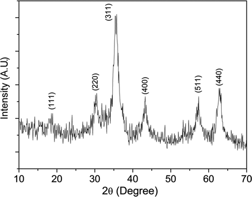
Figure 2. XRD patterns of the samples CoFe2− x Ho x O4 (x = 0.0, 0.025, 0.05, 0.075 and 0.1) annealed at 600°C.
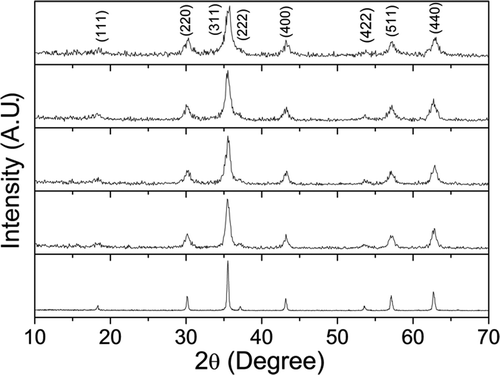
Figure 3. Rietveld refined XRD pattern for the sample CoFe1.95Ho0.05O4 annealed at 600°C. The circles represent experimental points and the solid line represents Rietveld refined data. The bottom line shows the difference between the experimental and refined data. The marked 2θ positions are the allowed Bragg peaks.
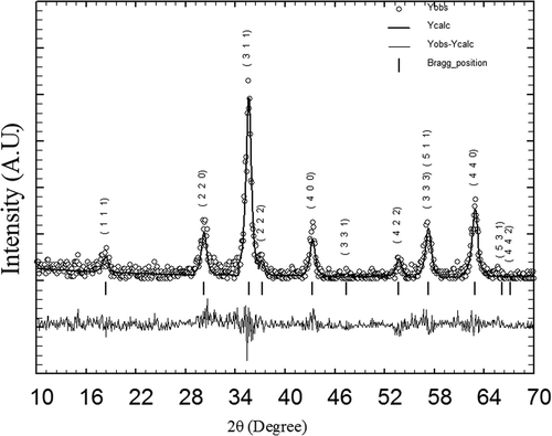
Table 1. Atomic coordinates and isothermal parameters for CoFe1.95Ho0.05O4 composition.
Table 2. Parameters obtained from Rietveld analysis of XRD patterns for the sample CoFe2− x Ho x O4 (x = 0.0, 0.025, 0.05, 0.075 and 0.1) annealed at 600°C. Errors of the lattice parameters are shown in brackets.
Table 3. Site occupancies of cations for the samples CoFe2− x Ho x O4 (x = 0.0, 0.025, 0.05, 0.075 and 0.1) annealed at 600°C.
Figure 4. FT-IR spectra of the samples CoFe2− x Ho x O4 with x = 0.0 and 0.025 annealed at 600°C.
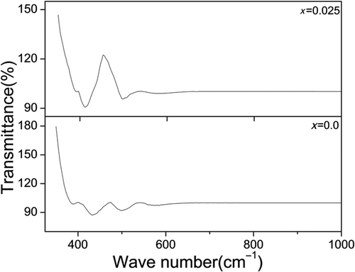
Figure 5. FE-SEM image of the sample CoFe1.975Ho0.025O4 annealed at 600°C.
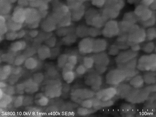
Figure 6. The EDS pattern of the sample CoFe1.975Ho0.025O4 annealed at 600°C. Au (gold) peaks are due to thin coating on the sample.
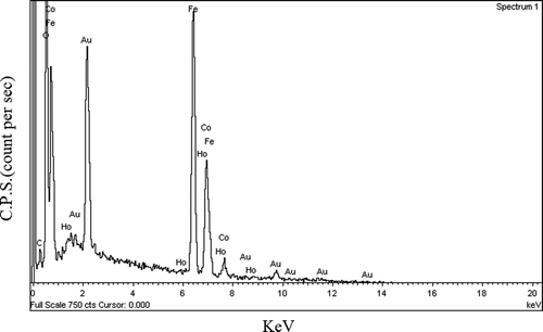
Figure 7. Hysteresis loops for the samples CoFe2− x Ho x O4 (x = 0.0, 0.025, 0.05, 0.075 and 0.1) annealed at 600°C.
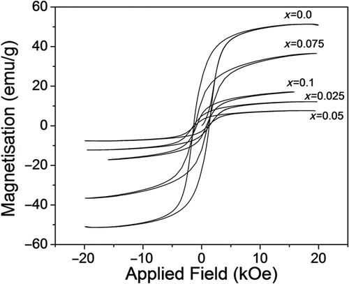
Table 4. Coercivity, saturation magnetisation (M S), and anisotropic constant (K 1) of the samples CoFe2− x Ho x O4 (x = 0, 0.025, 0.05, 0.075 and 0.1) annealed at 600°C.
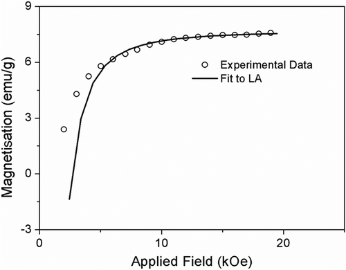
Figure 8. Fit to LA for the sample CoFe1.95Ho0.05O4 annealed at 600°C.