Figures & data
Figure 1. Scanning electron micrograph representing the shape and surface topology of the colloidal sulphur particles.
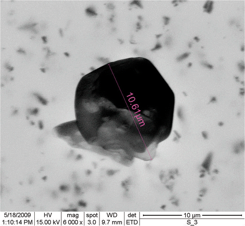
Figure 2. (A) Determination of actual (core) size of custom-made, (B) orthorhombic and (C) monoclinic SNPs.
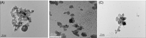
Table 1. The aggregation ratio characteristics of the adhesion between the SNPs in use
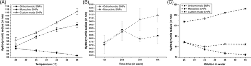
Figure 3. (A) Effect of temperature, (B) time drive (C) and dilution on the hydrodynamic radius of custom-made and synthesised SNPs.