
Figures & data
Figure 1. Spectroscopic studies: Ultraviolet-visible spectroscopic analysis of biologically synthesised gold nanoparticles (X-GNP) and chemically prepared gold nanoparticles (GNP).
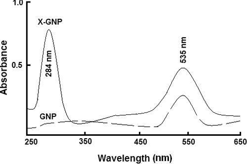
Figure 2. Size and charge distribution: Determination of size and surface charge of synthesised nanoparticles (GNP and X-GNP): (a) Dynamic light scattering (DLS) analysis of GNP and X-GNP suspension. (b) Estimation of surface charge of GNP and X-GNP by zeta potential measurement.
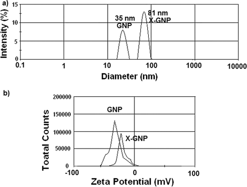
Figure 3. Microscopic study to determine the size and shape of the nanoparticles: Atomic force microscopic (AFM) images of GNP (a) and X-GNP (b). Scanning electron microscopy analysis was done for gold nanoparticles (c) and phytochemical-tagged gold nanoparticles (X-GNP).
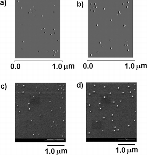
Figure 4. Elemental analysis: EDX analysis was done for both GNP (a) and X-GNP (b) to determine elemental components of synthesised nanoparticles.
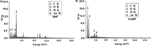
Figure 5. Characteristic Fourier-transform infrared absorption (FT-IR) spectra for GNP and biologically synthesised X-GNP.
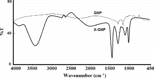
Figure 6. Antimicrobial activity: Growth inhibition percentages of five MTCC strains in the presence of (a) GNP and (b) X-GNP at different concentrations in broth assay.
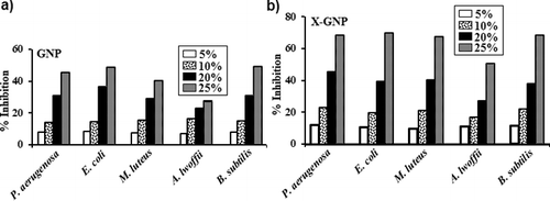
Figure 7. Batch growth profiles of P. aeruginosa (a), E. coli (b), M. luteus (c), A. lwoffii (d) and B. subtilis (e) with 25% supplements of GNP and X-GNP. NB (Nutrient broth, pH 7.0) with fraction-X was taken as (+) ve control and NB without any supplement was taken as (-) ve control. The maximum percentage of growth inhibition (f) with 25% supplements of fraction-X, GNP and X-GNP was plotted after 24 h of incubation at 37 °C.
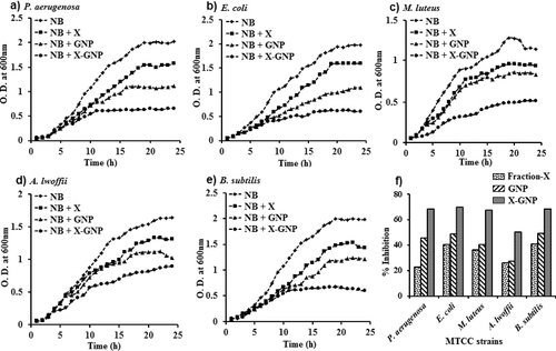
Table 1. Efficacy of GNP and X-GNP expressed as the zone of inhibition in spread plate cotton cloth diffusion studies.