Figures & data
Figure 1. Electrical scheme of the coupling between a neuron and a field effect transistor (adapted from Schatzthauer and Fromherz Citation27).

Figure 2. Schematical representation of the chemistry used to couple the PA22-2 peptide on the 16 MHA.
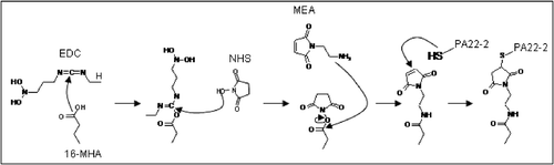
Figure 3. (a) Grazing Angle FTIR spectrum of PA22-2 immobilisation on a gold surface. Upon coupling of the peptide a large increase in the amide band and the hydroxyl band between 3000 cm−1 and 3500 cm−1 is observed as well as a reappearance of the carboxyl band at 1610 cm−1, (b) Enlarged part of the Grazing Angle FTIR spectrum of PA22-2 immobilisation on a gold surface. The shift of the carboxyl band from 1610 cm−1 to the maleimide band at 1750 cm−1 upon coupling the MEA can be observed.
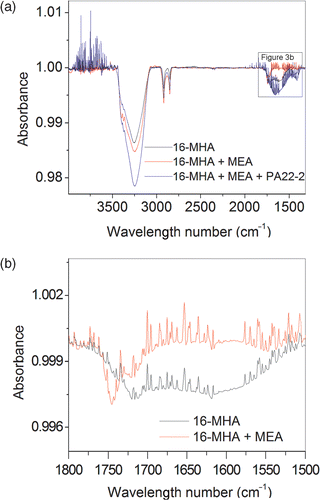
Figure 4. Comparable cultures of hippocampal neurons after 14 days in culture on PA22-2 (a) and PLL (b) coated gold substrates. More neurites are apparent on the PA22-2 coated surface.
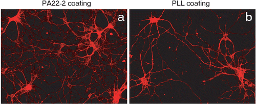
Figure 5. Proliferation results obtained with the Alamar Blue assay performed with Neuro-2a cells on differently coated substrate. The substrate coated with PA22-2 obtains similar results compared to PLL but is significantly better in sustaining the proliferation compared to bare gold substrates or 16 MHA coated substrates.
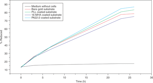
Figure 6. FTIR spectrum of microbeads coated with PA22-2 compared to control beads. The amide band appearing around 3500 cm−1 is typical for the presence of peptide on the beads.
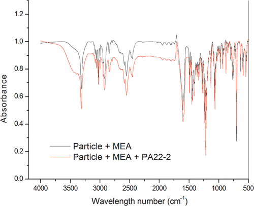
Figure 7. Interaction of HeLa cells with microbeads. (a) and (c) are control HeLa cells, (b) and (d) are HeLa cells stably transfected with full length TLN. The uncoated beads (a) and (b) are scattered in between the cells. The PA22-2 coated beads (c) and (d) bind to control HeLa cells and with more preference to the stable TLN expressing line.
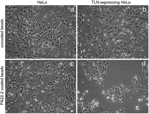
Figure 8. Higher magnification of the interaction between coated beads and transiently transfected HeLa Cells (a and b) and 7 days old primary culture of neurons (c and d). After incubation with the coated microbeads (green) and staining for TLN (red) and actin and nucleus (blue), it is shown that the beads are enriched on the TLN positive cells in panels a and b and that the coated beads redistribute along the neurites as observed in panel c and d.
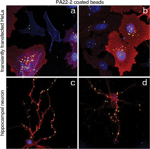
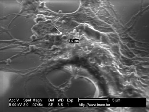
Figure 9. SEM picture of the neuronal cell membrane engulfing PA22-2 coated particles. The arrow points towards an apparently partial engulfed particle.