Figures & data
Figure 1. The figure represents measurable effective voxel of volume 1000R 3 containing superparamagnetic sphere of radius R in the center. Due to dephasing, it creates signal loss as MRI visible contrast in the ten fold boxed region (10R) or 1000R 3MRI visible volume of interest.
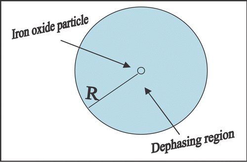
Figure 2. A mouse was injected with gadolinium bund MION-myoglobin particles (courtesy of Dr. Yousef Haik) through a vein in its tail and imaged by MRI at 500 MHz. The vascular regions showed clear wall and lumen areas as shown by arrows. Multislice-multiecho (Bruker Biospin at TE = 15 ms, TR = 1500 ms) gave proton density T 2* images with spatial resolution of 100 microns.
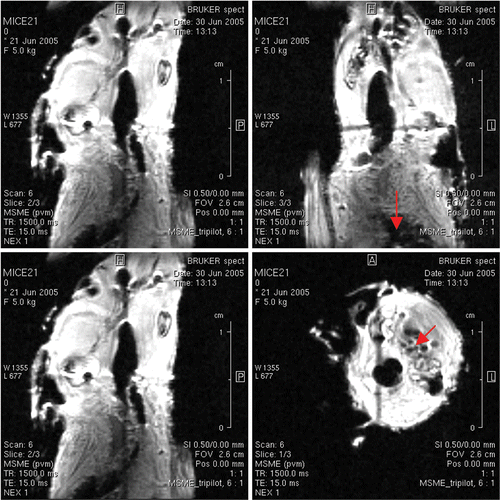
Figure 3. Quantitative T1 maps with pseudocolour obtained from solutions imaged in glass MR tubes. The tubes demonstrate a marked T1 shortening effect by loading GdHPDO3A (Prohance) into apoferritin. The sample on the lower right contains 100 mM Prohance only. After loading, the apoferritin will be extensively dialysed to remove free Prohance from solution, ensuring that the observed increase in relaxivity will be due to GDIII-apoferritin complexes.
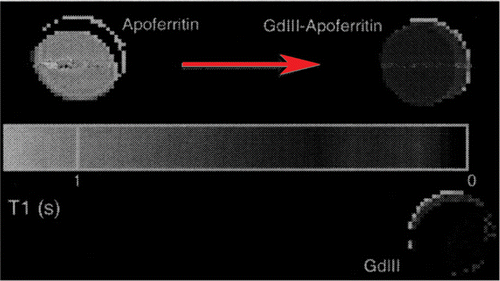
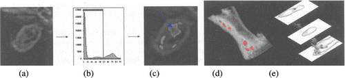
Figure 4. Axial section of the brachiocephalic artery image in control shown in panel (a) and after ‘artery injury’ induced mice ex vivo showing lipid rich necrotic core enhancement by iron oxide particles (arrow in c). Plaque components can be quantified using semi-automated segmentation techniques based on signal intensity histograms shown in panel (b). Segmentation allows quantification of individual vessel components and, in addition, (c) the segmented images can discriminate vessel damage shown with arrow in panel c. Combining slices, it can be reconstructed to show anatomical relationships of different components in 3D such as (demarcated necrotic cores in d). Wall details are visible in three different levels (e).