725
Views
21
CrossRef citations to date
0
Altmetric
Original Articles

Tuning hierarchical architecture of 3D polymeric scaffolds for cardiac tissue engineering

, , , , , , , , , , & show all
Pages 97-110 | Received 01 Aug 2007, Accepted 29 Sep 2007, Published online: 13 Aug 2008

Figures & data

Figure 1. Different length scales of cardiac tissue to be integrated into the scaffold: mm scale for vascularisation and nutrition, µm scale for cell accomodation, nm scale for the expression of ECM components.

Figure 1. Different length scales of cardiac tissue to be integrated into the scaffold: mm scale for vascularisation and nutrition, µm scale for cell accomodation, nm scale for the expression of ECM components.

Figure 2. SEM micrographs of PLA scaffolds prepared by: (a) NaCl leaching technique, the inset reports a zoomed area of the scaffold; (b) paraffin leaching techniques, the inset reports the cross-sectional view.
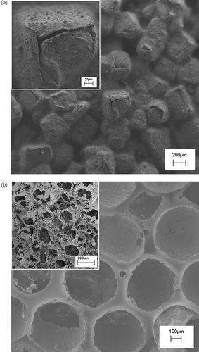
Figure 2. SEM micrographs of PLA scaffolds prepared by: (a) NaCl leaching technique, the inset reports a zoomed area of the scaffold; (b) paraffin leaching techniques, the inset reports the cross-sectional view.

Figure 3. SEM images of a PLA scaffold prepared by the phase separation technique at two different temperature (T) of solvent extraction, (a) T = −18°C and (b) −18°C < T < 25°C. The insets show high magnification SEM micrographs of a PLA scaffold prepared by the phase separation technique at two different temperature (T) of solvent extraction, (a) T 1 = −18°C and (b) −18°C < T 2 < 25°C.
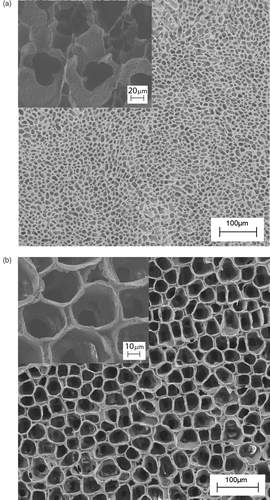
Figure 3. SEM images of a PLA scaffold prepared by the phase separation technique at two different temperature (T) of solvent extraction, (a) T = −18°C and (b) −18°C < T < 25°C. The insets show high magnification SEM micrographs of a PLA scaffold prepared by the phase separation technique at two different temperature (T) of solvent extraction, (a) T 1 = −18°C and (b) −18°C < T 2 < 25°C.

Figure 4. SEM image of PLA electrospun nanofibers with nanopores.

Figure 4. SEM image of PLA electrospun nanofibers with nanopores.

Figure 5. Schematics of the electrospinning experimental set up, where the grounded collecting electrodes were (a) two Cu parallel bars, (b) two pairs of Cu bar electrodes placed orthogonal to each other.
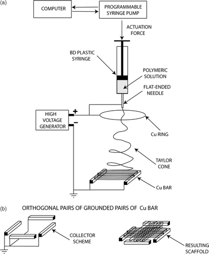
Figure 5. Schematics of the electrospinning experimental set up, where the grounded collecting electrodes were (a) two Cu parallel bars, (b) two pairs of Cu bar electrodes placed orthogonal to each other.

Figure 6. SEM micrographs of (a) oriented PCL fiber bundle, (b) macroscopically textured PCL scaffold.
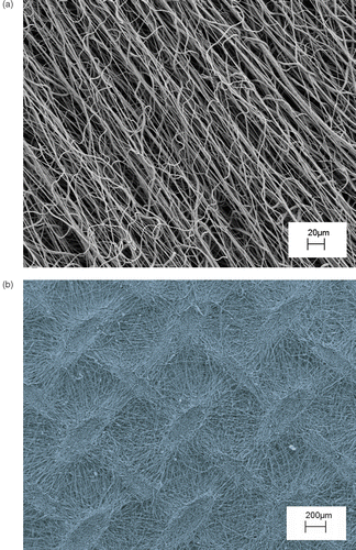
Figure 6. SEM micrographs of (a) oriented PCL fiber bundle, (b) macroscopically textured PCL scaffold.

Figure 7. Immunofluorescence analysis of F-actin expression (red) in mMSC grown after 72 h, on (a) PLA scaffold shown in , (b) PCL scaffold shown in . Nuclei were stained with 4′,6′-diamidino-2-phenylindole (DAPI). Images of cell samples were taken with a Leica DMRB microscope using a digital camera. The images were representative of at least five random fields for each sample.
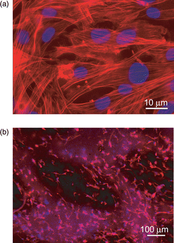
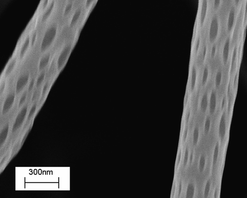
Figure 7. Immunofluorescence analysis of F-actin expression (red) in mMSC grown after 72 h, on (a) PLA scaffold shown in Figure 3(b), (b) PCL scaffold shown in Figure 6(b). Nuclei were stained with 4′,6′-diamidino-2-phenylindole (DAPI). Images of cell samples were taken with a Leica DMRB microscope using a digital camera. The images were representative of at least five random fields for each sample.

Reprints and Corporate Permissions

Please note: Selecting permissions does not provide access to the full text of the article, please see our help page How do I view content?

To request a reprint or corporate permissions for this article, please click on the relevant link below:

Academic Permissions

Please note: Selecting permissions does not provide access to the full text of the article, please see our help page How do I view content?

Obtain permissions instantly via Rightslink by clicking on the button below:

If you are unable to obtain permissions via Rightslink, please complete and submit this Permissions form. For more information, please visit our Permissions help page.