549
Views
6
CrossRef citations to date
0
Altmetric
Original Articles

Functionalised microneedles for enhanced neuronal adhesion

, , , , , , , , & show all
Pages 147-156 | Received 27 Jun 2007, Accepted 12 Mar 2008, Published online: 13 Aug 2008

Figures & data

Figure 1. Schematic comparison of a neuron–sensor interface using a flat sensor and a sensor with needles.

Figure 1. Schematic comparison of a neuron–sensor interface using a flat sensor and a sensor with needles.

Figure 2. Schematic representation of the PA22-2 immobilisation on gold surfaces.

Figure 2. Schematic representation of the PA22-2 immobilisation on gold surfaces.

Figure 3. SEM picture: (a) an array of Au needles and (b) detailed view of a single needle.

Figure 3. SEM picture: (a) an array of Au needles and (b) detailed view of a single needle.

Figure 4. Coupling of the peptide on the gold needles of the chip: (a) brightfield image of the chip and (b) fluorescent image of the same area.
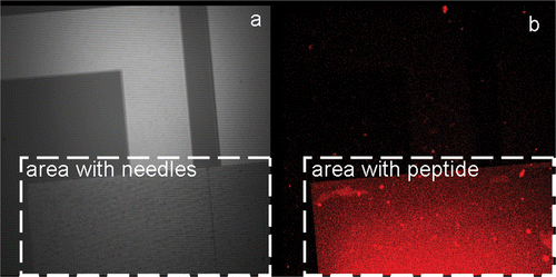
Figure 4. Coupling of the peptide on the gold needles of the chip: (a) brightfield image of the chip and (b) fluorescent image of the same area.

Figure 5. Z-stack of TLN expressing HeLa cells cultured on top of Alexa 555 labelled PA22-2 on top of the needles. The small insets form a schematic representation of the experimental set-up with the confocal plane of the picture as a dashed line. The consecutive pictures are taken at 0.5 µm intervals.
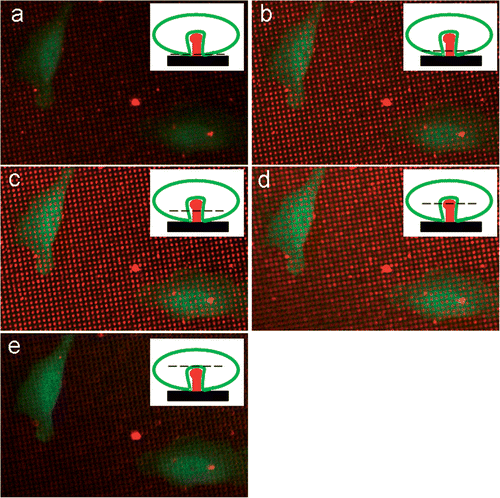
Figure 5. Z-stack of TLN expressing HeLa cells cultured on top of Alexa 555 labelled PA22-2 on top of the needles. The small insets form a schematic representation of the experimental set-up with the confocal plane of the picture as a dashed line. The consecutive pictures are taken at 0.5 µm intervals.
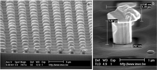
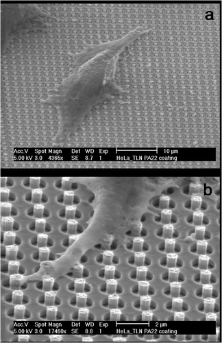
Figure 6. SEM pictures of HeLa cells growing on needles: (a) overview picture of a cell growing on a needle-bed. (b) Detailed picture of a cell extension that engulfs several needles.

Figure 6. SEM pictures of HeLa cells growing on needles: (a) overview picture of a cell growing on a needle-bed. (b) Detailed picture of a cell extension that engulfs several needles.

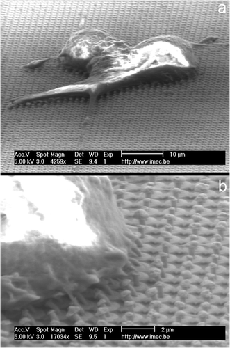
Figure 7. SEM pictures of PC12 cells growing on PA22-2-coated gold needles: (a) overview of the entire cell as it grows on the needles. (b) Detailed view showing a strong interaction between the cell membrane and the needles that becomes visible through the bending of the needles.

Figure 7. SEM pictures of PC12 cells growing on PA22-2-coated gold needles: (a) overview of the entire cell as it grows on the needles. (b) Detailed view showing a strong interaction between the cell membrane and the needles that becomes visible through the bending of the needles.

Reprints and Corporate Permissions

Please note: Selecting permissions does not provide access to the full text of the article, please see our help page How do I view content?

To request a reprint or corporate permissions for this article, please click on the relevant link below:

Academic Permissions

Please note: Selecting permissions does not provide access to the full text of the article, please see our help page How do I view content?

Obtain permissions instantly via Rightslink by clicking on the button below:

If you are unable to obtain permissions via Rightslink, please complete and submit this Permissions form. For more information, please visit our Permissions help page.