Figures & data
Figure 1. IR spectraof the (a) as-obtained, (b) amide-derivatised, (c) HDTMS-coated, (d) DBDMT-coated, (e) PFOTES-coated, (f) silica-coated and (g) tin oxide-coated nanodiamond.
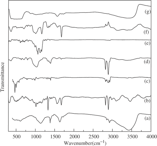
Figure 2. TEM images of the (a) as-obtained nanodiamond with the inset showing the particle size distribution, (b) amide-derivatised nanodiamond with the EDAX pattern as the inset, (c) HDTMS-coated nanodiamond with the EDAX pattern as the inset and (d) DBDMT-coated nanodiamond with the EDAX patter as the inset.
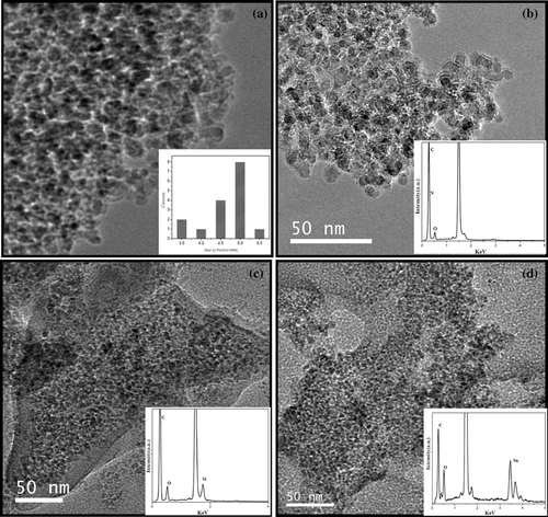
Figure 3. Photographs of dispersions of (a) amide-derivatised, (b) HDTMS-coated (c) PFOTES-coated and (d) DBDMT-coated nanodiamonds in nonpolar solvents.
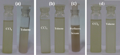
Figure 4. Photographs of dispersions of nanodiamond induced by (a) IGEPAL, (b) CTAB, (c) TX-100, (d) PVA and (e) AOT in water and (f) AOT in toluene.
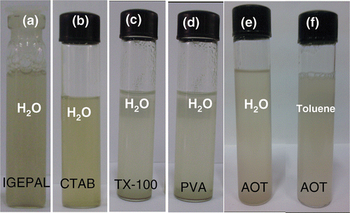
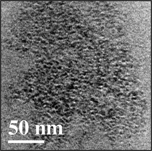
Figure 5. TEM image of the PMMA-nanodiamond composite.