637
Views
3
CrossRef citations to date
0
Altmetric
Original Articles

Synthesis of nano-hydroxyapatite from silica suspensions through chemical compensation

, &
Pages 193-202 | Received 08 Dec 2008, Accepted 23 Jan 2009, Published online: 22 May 2009

Figures & data

Figure 1. SEM micrograph of sample prepared as described in the text at pH = 6. Hap needles about 1 µm long and 0.1 µm thick form bundles from a common origin. These bundles join one another and fill the space (marker = 1 µm).
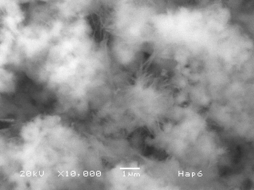
Figure 1. SEM micrograph of sample prepared as described in the text at pH = 6. Hap needles about 1 µm long and 0.1 µm thick form bundles from a common origin. These bundles join one another and fill the space (marker = 1 µm).

Figure 2. XRD from sample in shows the presence of Hap with very high crystallinity. Peaks for other calcium phosphates are not present.
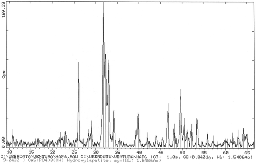
Figure 2. XRD from sample in Figure 1 shows the presence of Hap with very high crystallinity. Peaks for other calcium phosphates are not present.

Figure 3. SEM micrograph of sample prepared at pH = 9. Hap needles 12 µm long are observed, some as thick as 1 µm are present. Some form bundles from a common origin.
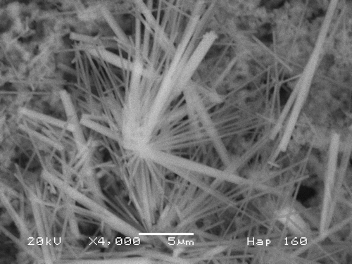
Figure 3. SEM micrograph of sample prepared at pH = 9. Hap needles 12 µm long are observed, some as thick as 1 µm are present. Some form bundles from a common origin.

Figure 4. XRD of sample of shows the presence of Hap with very high crystallinity. No peaks for other calcium phosphates are present.
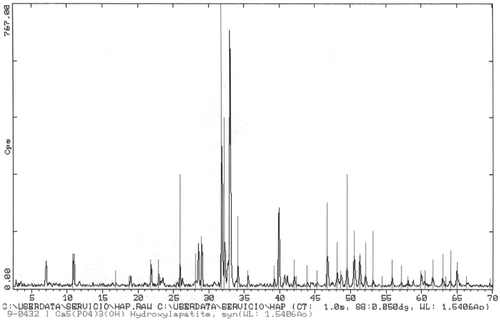
Figure 4. XRD of sample of Figure 3 shows the presence of Hap with very high crystallinity. No peaks for other calcium phosphates are present.

Figure 5. Backscattered electrons micrograph from sample prepared at pH = 6 shows elongated crystallites about 5 µm long and 1 µm thick embedded in a SiO2 matrix. Hap crystallites appear isolated, not in bundles.
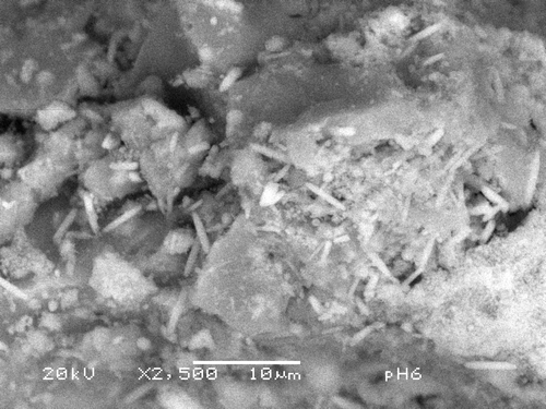
Figure 5. Backscattered electrons micrograph from sample prepared at pH = 6 shows elongated crystallites about 5 µm long and 1 µm thick embedded in a SiO2 matrix. Hap crystallites appear isolated, not in bundles.

Figure 6. XRD from sample in . It shows that the needles are crystalline Hap, although a couple of extra peaks are present. These could be due to CaF2 or KCl2.
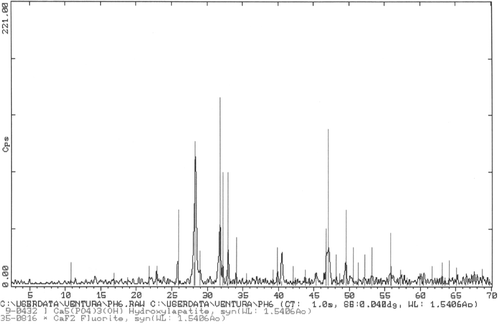
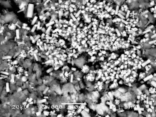
Figure 6. XRD from sample in Figure 5. It shows that the needles are crystalline Hap, although a couple of extra peaks are present. These could be due to CaF2 or KCl2.
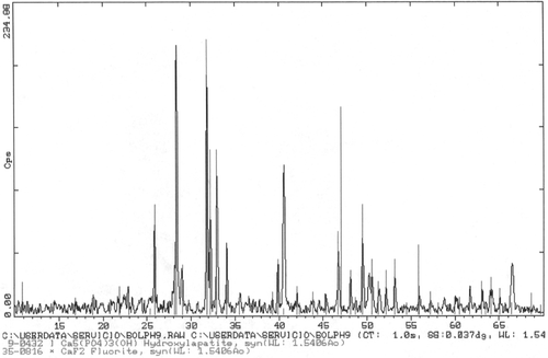
Figure 7. SEM micrograph of sample prepared as in Section 2.2.2 at pH = 9. Hap bars are observed as growing on top of the SiO2 particles. Bars are about 8 µm × 1 µm and SiO2 particles show dimensions in the order of 5 µm. The hexagonal cross section is clearly observed.

Figure 7. SEM micrograph of sample prepared as in Section 2.2.2 at pH = 9. Hap bars are observed as growing on top of the SiO2 particles. Bars are about 8 µm × 1 µm and SiO2 particles show dimensions in the order of 5 µm. The hexagonal cross section is clearly observed.

Figure 8. XRD from sample in . It shows that the bars correspond to crystalline Hap, although other peaks are present. These could be due to CaF2 or KCl2.

Figure 8. XRD from sample in Figure 7. It shows that the bars correspond to crystalline Hap, although other peaks are present. These could be due to CaF2 or KCl2.

Reprints and Corporate Permissions

Please note: Selecting permissions does not provide access to the full text of the article, please see our help page How do I view content?

To request a reprint or corporate permissions for this article, please click on the relevant link below:

Academic Permissions

Please note: Selecting permissions does not provide access to the full text of the article, please see our help page How do I view content?

Obtain permissions instantly via Rightslink by clicking on the button below:

If you are unable to obtain permissions via Rightslink, please complete and submit this Permissions form. For more information, please visit our Permissions help page.