8,676
Views
32
CrossRef citations to date
0
Altmetric
Review

Novel CNS drug discovery and development approach: model-based integration to predict neuro-pharmacokinetics and pharmacodynamics

, , , & ORCID Icon
Pages 1207-1218 | Received 20 Jun 2017, Accepted 13 Sep 2017, Published online: 21 Sep 2017

Figures & data

Figure 1. The combinatory mapping approach. (Redrawn from [Citation23] with permission of Springer).

Figure 1. The combinatory mapping approach. (Redrawn from [Citation23] with permission of Springer).

Figure 2. The structure of the generic CNS PBPK model Structure: Black: Plasma PK model, Red: CNS PBPK model. Parameters: Black: estimated plasma PK parameters; Blue: system-specific parameters; Green: drug-specific parameters; Purple: combination of system-specific and drug-specific specific parameters. (Redrawn from [Citation39] with permission of Wiley & Sons).

Figure 2. The structure of the generic CNS PBPK model Structure: Black: Plasma PK model, Red: CNS PBPK model. Parameters: Black: estimated plasma PK parameters; Blue: system-specific parameters; Green: drug-specific parameters; Purple: combination of system-specific and drug-specific specific parameters. (Redrawn from [Citation39] with permission of Wiley & Sons).

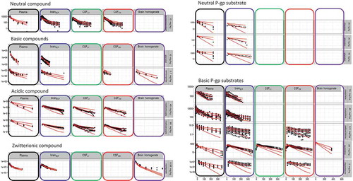
Figure 3. BPPK model prediction (red lines for median and 95% percentiles) and observed data (black dots) obtained in the five color-filled compartments.

Figure 3. BPPK model prediction (red lines for median and 95% percentiles) and observed data (black dots) obtained in the five color-filled compartments.

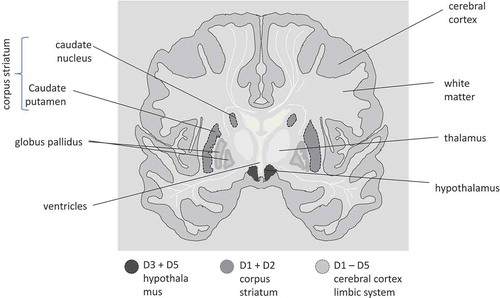
Figure 4. Cartoon of transversal cross section of human brain with the dopaminergic receptor areas indicated. Receptor density is region specific and receptor subtype specific.

Figure 4. Cartoon of transversal cross section of human brain with the dopaminergic receptor areas indicated. Receptor density is region specific and receptor subtype specific.

Figure 5. Compared plasma pharmacokinetics of high affinity compounds (top row) and their low affinity analogues (bottom row). The circles represent observed plasma concentrations in rats, the lines represent model predictions. (Reprinted from [Citation45] with permission of ASPET).

Figure 5. Compared plasma pharmacokinetics of high affinity compounds (top row) and their low affinity analogues (bottom row). The circles represent observed plasma concentrations in rats, the lines represent model predictions. (Reprinted from [Citation45] with permission of ASPET).

Table 1. Biomarker classification [Citation71] and approaches to assess quantitative information.