Figures & data
Figure 1. Schematic representation of: (i) the sheared process used to fabricate fluid gels. Reproduced from [Citation29], licensed with CC BY 4.0 (https://creativecommons.org/licenses/by/4.0/). (ii) The microstructure of the fluid gel (particles suspended in interstitial polymer. Reproduced from [Citation30], licensed with CC BY 4.0 (https://creativecommons.org/licenses/by/4.0/). (iii) Representation of interactions between the particles and polymer phase. Reproduced from [Citation30], licensed with CC BY 4.0 (https://creativecommons.org/licenses/by/4.0/).
![Figure 1. Schematic representation of: (i) the sheared process used to fabricate fluid gels. Reproduced from [Citation29], licensed with CC BY 4.0 (https://creativecommons.org/licenses/by/4.0/). (ii) The microstructure of the fluid gel (particles suspended in interstitial polymer. Reproduced from [Citation30], licensed with CC BY 4.0 (https://creativecommons.org/licenses/by/4.0/). (iii) Representation of interactions between the particles and polymer phase. Reproduced from [Citation30], licensed with CC BY 4.0 (https://creativecommons.org/licenses/by/4.0/).](/cms/asset/9b295e2b-f691-45a2-86e9-ead07489ab82/ierl_a_2101998_f0001_oc.jpg)
Figure 2. (a) Mechanical analysis showing transition from liquid to solid as the eye drop it is squeezed from the applicator. Reproduced from [Citation29], licensed with CC BY 4.0 (https://creativecommons.org/licenses/by/4.0/). (b) Preclinical retention data obtained on a rat model showing retention over several hours. Reproduced from [Citation42], licensed with CC BY 4.0 (https://creativecommons.org/licenses/by/4.0/). (c) Role of lubrication throughout the blinking process: (i) Schematic representation of Stribeck curve highlighting boundary, mixed and hydrodynamic regimes; (ii) Tribology data for fluid gels showing the reduction in friction coefficient (lubricity) as a function of the formulation. Reproduced from [Citation35], licensed with CC BY 3.0 (https://creativecommons.org/licenses/by/3.0/).
![Figure 2. (a) Mechanical analysis showing transition from liquid to solid as the eye drop it is squeezed from the applicator. Reproduced from [Citation29], licensed with CC BY 4.0 (https://creativecommons.org/licenses/by/4.0/). (b) Preclinical retention data obtained on a rat model showing retention over several hours. Reproduced from [Citation42], licensed with CC BY 4.0 (https://creativecommons.org/licenses/by/4.0/). (c) Role of lubrication throughout the blinking process: (i) Schematic representation of Stribeck curve highlighting boundary, mixed and hydrodynamic regimes; (ii) Tribology data for fluid gels showing the reduction in friction coefficient (lubricity) as a function of the formulation. Reproduced from [Citation35], licensed with CC BY 3.0 (https://creativecommons.org/licenses/by/3.0/).](/cms/asset/474fa1d1-2e85-43e5-8a60-662b78ee0d69/ierl_a_2101998_f0002_oc.jpg)
Figure 3. (a) Macro and histological images of the cornea following various treatments including standard of care (group 1), standard of care with fluid gel (group 2) and standard of care with fluid gel loaded with decorin (group 3) in a model of pseudomonas keratitis. (b) Immunohistochemistry of the cornea having been stained for specific fibrotic markers: (i)αSMA; (ii) Fibronectin; (iii) Laminin. Data show that fluid gel with standard care (group 2) is superior to standard care alone (group 1), while FG with decorin (group 3) restores the cornea to an ‘intact’ baseline phenotype 16 days post-treatment. Reproduced from [Citation29], licensed with CC BY 4.0 (https://creativecommons.org/licenses/by/4.0/).
![Figure 3. (a) Macro and histological images of the cornea following various treatments including standard of care (group 1), standard of care with fluid gel (group 2) and standard of care with fluid gel loaded with decorin (group 3) in a model of pseudomonas keratitis. (b) Immunohistochemistry of the cornea having been stained for specific fibrotic markers: (i)αSMA; (ii) Fibronectin; (iii) Laminin. Data show that fluid gel with standard care (group 2) is superior to standard care alone (group 1), while FG with decorin (group 3) restores the cornea to an ‘intact’ baseline phenotype 16 days post-treatment. Reproduced from [Citation29], licensed with CC BY 4.0 (https://creativecommons.org/licenses/by/4.0/).](/cms/asset/8385e7c8-c949-4241-bae0-a0d2d520e165/ierl_a_2101998_f0003_oc.jpg)
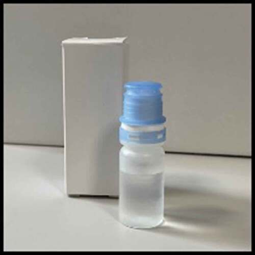
Figure 4. Schematic representation of the fluid gel eye drop upon the cornea, highlighting how the material will aid as an osmoprotectant, by providing tear film retention value through structuring ‘trapping’ of the water. Its hydrophilic nature (potentially) overcomes the loss of mucin seen in dry eye disease: thereby FG technology will go a long way to recapitulating the tear film microarchitecture.
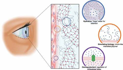
Figure 5. Image of the fluid gel eye drop in packaging ready for Phase I clinical trials.