Figures & data
Figure 1. Ultrasound biomicroscopic (UBM) images illustrating how ALPI compacts the peripheral iris stroma, creating a space between the anterior iris surface and the trabecular meshwork, thus opening the angle. Left: appositionally closed angle in an eye with plateau iris syndrome (S: sclera, CB: ciliary body, AC: anterior chamber, C: cornea). Right: open angle after ALPI.
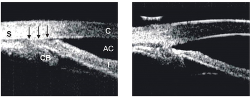
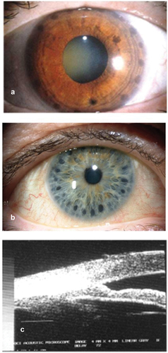
Figure 2. (a) Slit lamp photograph of an eye with plateau iris syndrome after ALPI. The dark, round laser marks can be clearly seen on the peripheral iris. (b) ALPI burns being placed too centrally, and thus ineffective. (c) UBM showing ALPI being placed too centrally, and the angle remains closed.
Reprinted from Survey of Ophthalmology, Vol 52, No. 3, Ritch R, Tham CC, Lam DS. Argon laser peripheral iridoplasty (ALPI): an update. P. 279–288, 2007, with permission from Elsevier.