Figures & data
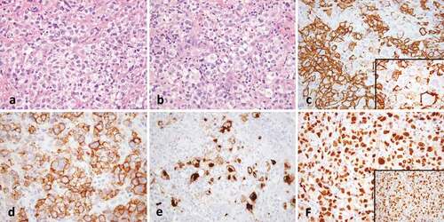
Figure 1. Gray-zone lymphoma. (a) There is a diffuse infiltrate of atypical lymphoid cells with clear cytoplasm (40x). (b) Occasional cells have Hodgkin-like features (40x). (c) CD20 shows strong staining with some variability (40x, inset 100x). (d) CD30 is strongly positive and (e) CD15 shows golgi and membranous staining in a subset of Hodgkin-like cells (both 20x). (f) OCT2 (40x) and PAX-5 (inset, 40x) are strongly positive.