
Figures & data
Figure 1. Targeting BCL-2 in MDS. (a) Anti- and pro-apoptotic members of the BCL-2 family proteins as regulators of the mitochondrial pathway of apoptosis. (b) Cancer cell survival: BCL-2 overexpression sequestering high levels of pro-apoptotic proteins prevents cells from initiating apoptosis. (c) Induction of apoptosis: displacing BIM and BAK by venetoclax allows pro-apoptotic proteins to initiate apoptosis. (d) Suggested synergy between hypomethylating agents and venetoclax. (e) Decreasing basal apoptosis rate with increasing BCL-2 dependency and suggested higher venetoclax sensitivity upon disease progression in MDS
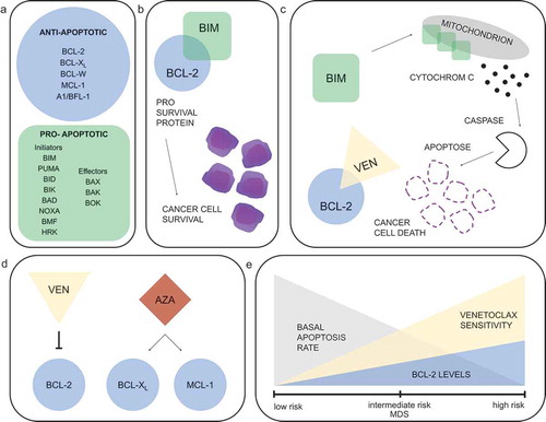
Table 1. Selection of current clinical trials with venetoclax in MDS
Table 2. Selection of clinical trials with venetoclax in myeloid malignancies including MDS