Figures & data
Table 1. Prevalence and type of fetal anemia for different types of MC twin complications.
Figure 1. (a) Sonographic image of placental dichotomy, with hyperechogenic hydropic placental share for the TAPS donor and flattened hypoechogenic placental share for the TAPS recipient. (b) Cardiomegaly in the TAPS donor. (c) Starry-sky liver in the TAPS recipient.
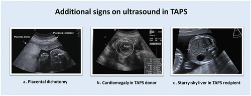
Table 2. Antenatal classification system for TAPS.
Figure 2. A MC twin pair with spontaneous twin anemia polycythemia sequence illustrating the characteristic large skin color difference.
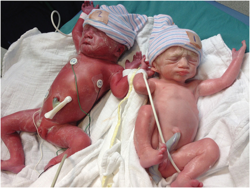
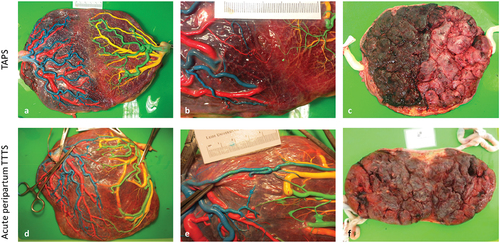
Figure 3. An overview of placental differences between TAPS (upper row) and acute peripartum TTTS (lower row). Placentas have been injected with colored dye. Blue (arteries) and red (veins) were used for the first-born twin and green (arteries) and yellow (veins) were used for the second-born twin. (a) Fetal side of the a placenta from spontaneous TAPS. The left placental share belongs to the recipient twin, the right placental share belongs to the donor twin. (b) Color dye injection revealed only one minuscule veno-arterial anastomosis (red-green). (c) The maternal side of the TAPS placenta shows a striking color difference with a plethoric share for the recipient and a pale share for the donor. (d) Fetal side of a placenta from a acute peripartum TTTS twin. The left placental share belongs to the recipient twin, the left placental share to the donor twin. (e) Color dye injection revealed numerous large anastomoses including an arterio-arterial anastomosis (blue-green). (f) The maternal side of the acute peripartum TTTS placenta shows no color difference.