8,204
Views
17
CrossRef citations to date
0
Altmetric
Review

Utilizing artificial intelligence in endoscopy: a clinician’s guide

, , , , , , , , , & show all
Pages 689-706 | Received 31 Mar 2020, Accepted 03 Jun 2020, Published online: 17 Jun 2020

Figures & data

Figure 1. Representative images of gastric cancer detected by artificial intelligence.

(a) A reddish and slightly depressed lesion of gastric cancer appears on the greater curvature of the lower gastric body.(b) The yellow rectangular frame was marked by artificial intelligence as a possible lesion to indicate the extent of a suspected gastric cancer lesion. An endoscopist manually marked the location of the cancer using a green rectangular frame. [0–IIc, 10 mm, tub1, T1a(M)].
Figure 1. Representative images of gastric cancer detected by artificial intelligence.

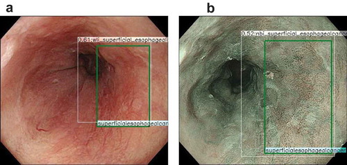
Figure 2. Representative images of esophageal cancer detected by artificial intelligence.

(a) Reddish irregular area of esophageal squamous cell carcinoma (ESCC) on the right wall in white light imaging. The endoscopist marked the lesion using a green square, and an artificial intelligence (AI) diagnosing system surrounded the lesion using a white square to diagnose esophageal cancer. Because both squares were matched perfectly, we know the AI diagnosing system could detect ESCC correctly.(b) In narrow band imaging, the same lesion appears as a Brownish area. The AI diagnosing system detected ESCC in the same way.
Figure 2. Representative images of esophageal cancer detected by artificial intelligence.

Table 1. Use of artificial intelligence in stomach field.

Table 2. Use of artificial intelligence in colonic polyp.

Table 3. Deep learning-based systems for capsule endoscopy.
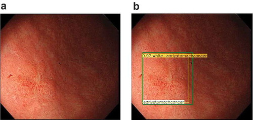
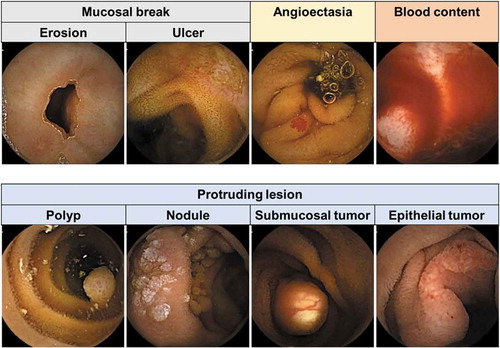
Figure 3. Representative images of various abnormalities captured by capsule endoscopy.

Figure 3. Representative images of various abnormalities captured by capsule endoscopy.