Figures & data
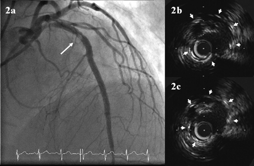
Figure 1. (a) Coronary angiography showing a stenosis with haziness in the proximal left anterior descending artery (LAD) two weeks after sirolimus-eluting stents (SES) implantation. (b) and (c) Intravascular ultrasound imaging of the stented segment at two weeks of follow-up. The maximal plaque prolapse cross sectional area was 2.5 mm2 with a percentage of plaque prolapse burden: 40% of stent area. Stent struts were labeled with arrows. (d) Coronary angiography showing partial resolution of the stenosis with haziness after balloon dilatation. (e) and (f) Intravascular ultrasound imaging of the stented segment after balloon dilatation.
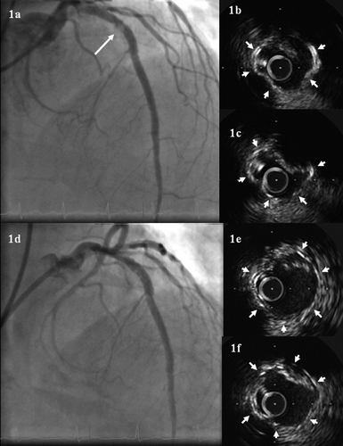
Figure 2. (a) Coronary angiography showing a similar stenosis with haziness in the proximal LAD six weeks after SES implantation. (b) and (c) Intravascular ultrasound imaging of the stented segment at six weeks of follow-up.