Figures & data
Figure 1. UV–Vis spectra of silver nanoparticles (PE-AgClNPs) synthesized by the peach outer peel extracts (PE). Inset: fruit and outer peel of peach (P. persica L.) (left); Change in color of the solution confirming the synthesis of PE-AgClNPs ((a) – AgNO3 solution, (b) – peach outer peel extracts (PE), (c) – PE-AgClNPs) (Right).
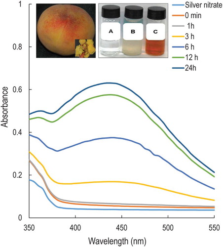
Figure 2. SEM image (a); size distribution graph (b) and EDX (c) of silver nanoparticles synthesized by the peach outer peel extracts.
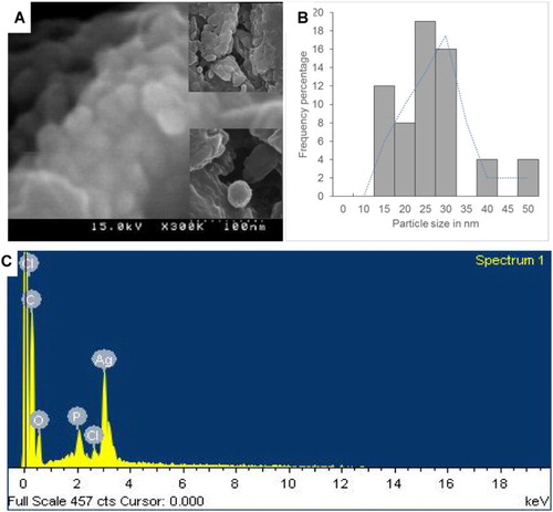
Figure 3. XRD (a), TGA (b; blue line is the TGA pattern of PE-AgClNPs and green line is the derivative weight of PE-AgClNPs) and FT-IR analysis (c) of silver nanoparticles (PE-AgClNPs) synthesized by the peach outer peel extracts (PE).
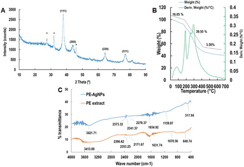
Table 1. Antibacterial activity of PE-AgClNPs (50 µg/disc) against five foodborne pathogenic bacteria.
Figure 4. Synergistic antibacterial potential and anticandidal activity of silver nanoparticles synthesized by the peach outer peel extracts. The green circles represent the diameter of zone of inhibition.
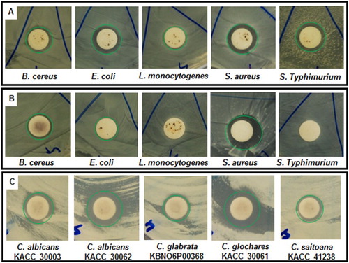
Table 2. Synergistic antibacterial activity of PE-AgClNPs (25 µg) with standard antibiotics, kanamycin (5 µg) or rifampicin (5 µg), against foodborne pathogenic bacteria.
Table 3. Synergistic anticandidal activity of PE-AgClNPs (50 µg) with standard antifungal agent, amphotericin b (5 µg), against pathogenic Candida species.
Figure 5. Antioxidant activity of silver nanoparticles synthesized by the peach outer peel extracts. DPPH free radical scavenging (A), nitric oxide scavenging (B), ABTS radical scavenging (C) and reducing power (D) potential of silver nanoparticles (PE-AgClNPs) and ascorbic acid as the reference compound. Different superscript letters in each column indicate significant differences at P < .05.