
Figures & data
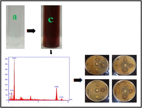
Figure 1. (a) Copper sulfate solution, (b) F. macrocolea flowers extract, (c) CuNPs colloidal suspension.
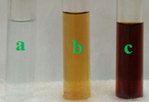
Figure 2. UV-Vis spectra of CuNPs synthesized by F. macrocolea flowers extract (a) Aqueous suspension, (b) Liquid ammonia suspension.
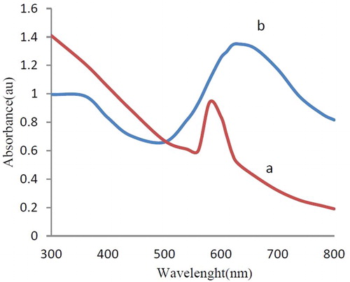
Figure 3. (a) FT-IR spectrum of F. macrocolea flowers aqueous extract. (b) FT-IR spectrum of the synthesized CuNPs.
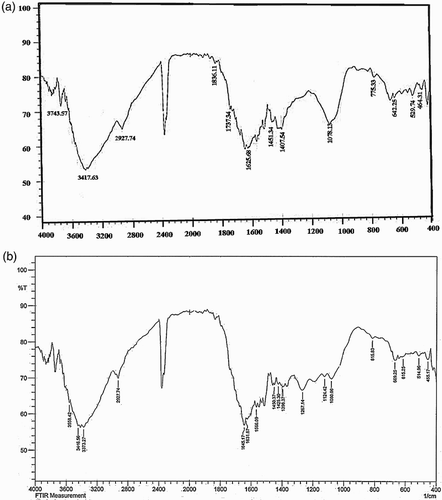
Figure 4. SEM image of the CuNPs.
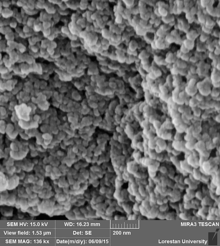
Figure 5. EDX image of the CuNPs.
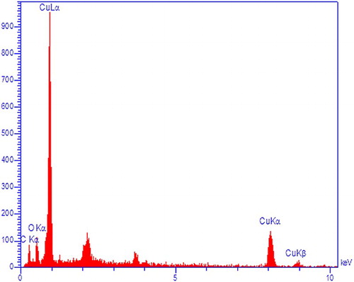
Figure 6. XRF image of the CuNPs.
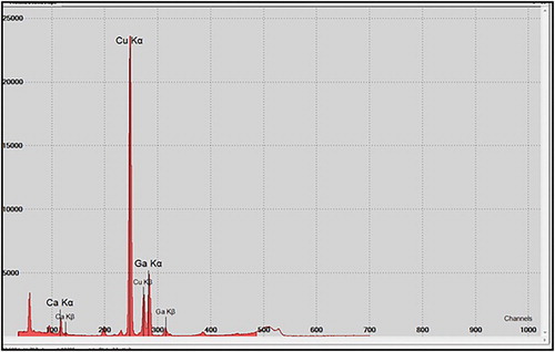
Figure 7. XRD pattern of the CuNPs.
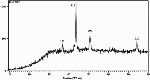
Figure 8. Antibacterial effect of the CuNPs against bacteria, (a) S. aureus, (b) B. cereus, (c) E. coli and (d) K. pneumonia.
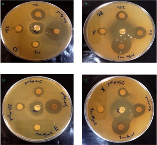
Table 1. Antibacterial activity of synthesized CuNPs.
Table 2. Percent viability of cells exposed to the CuNPs.
Supplemental material