9,396
Views
146
CrossRef citations to date
0
Altmetric
RESEARCH REVIEW

Anti-cancer green bionanomaterials: present status and future prospects

, , & ORCID Icon
Pages 285-314 | Received 27 Apr 2017, Accepted 25 Sep 2017, Published online: 20 Oct 2017

Figures & data

Figure 1. Biological synthesis and applications of metal NPs in biomedical and environmental fields (Adapted with permission from Singh et al. (Citation12)).

Figure 1. Biological synthesis and applications of metal NPs in biomedical and environmental fields (Adapted with permission from Singh et al. (Citation12)).

Figure 2. Schematic mechanism of microbial-mediated synthesis of metal nanoparticles (Adapted with permission from Salunke et al. (Citation11)).

Figure 2. Schematic mechanism of microbial-mediated synthesis of metal nanoparticles (Adapted with permission from Salunke et al. (Citation11)).

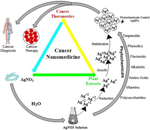
Figure 3. Plant-mediated mechanism of biogenic silver nanoparticles’ (AgNPs’) production: a step forward for cancer nanomedicine (Adapted with permission from Ovais et al. (Citation21)).

Figure 3. Plant-mediated mechanism of biogenic silver nanoparticles’ (AgNPs’) production: a step forward for cancer nanomedicine (Adapted with permission from Ovais et al. (Citation21)).

Table 1. Phyto-sources for synthesis of bionanoparticles and their anticancer activity in various cell lines for the past 5 years.
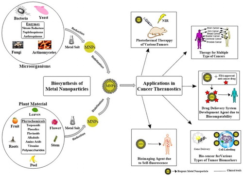
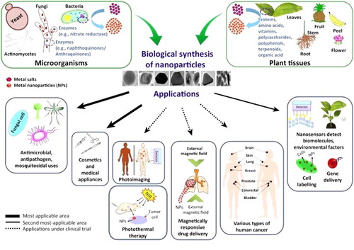
Table 2. Microbial sources for synthesis of bionanoparticles and their anticancer activity in various cell lines for the past 5 years.
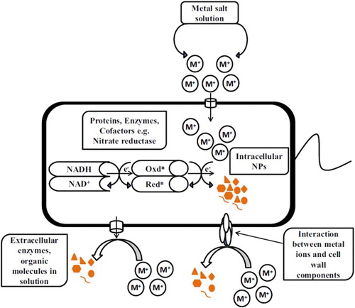
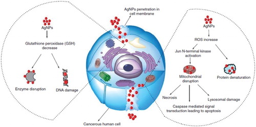
Figure 4. Recent advances in proposed mechanisms for anticancer activity shown by colloidal biogenic silver nanoparticles (AgNPs) (Adapted with permission from Ovais et al. (Citation21)).

Figure 4. Recent advances in proposed mechanisms for anticancer activity shown by colloidal biogenic silver nanoparticles (AgNPs) (Adapted with permission from Ovais et al. (Citation21)).

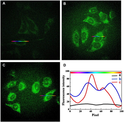
Figure 5. Fluorescence images of HeLa cancer cells which were incubated in the absence of Au/Ce NCs (A), in the presence of 50 μmol/L (B) and 150 μmol/L (C) Au/Ce NCs solutions for 24 h. (D) The fluorescence intensity variations along cross-sections a (in A), b (in B) or c (in C). Fluorescence images were collected by applying fluorescence excitation wavelength at 488 nm (Adapted with permission from Ge et al. (Citation118)).

Figure 5. Fluorescence images of HeLa cancer cells which were incubated in the absence of Au/Ce NCs (A), in the presence of 50 μmol/L (B) and 150 μmol/L (C) Au/Ce NCs solutions for 24 h. (D) The fluorescence intensity variations along cross-sections a (in A), b (in B) or c (in C). Fluorescence images were collected by applying fluorescence excitation wavelength at 488 nm (Adapted with permission from Ge et al. (Citation118)).

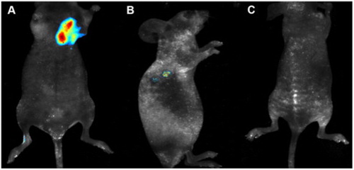
Figure 6. Tumor nude mice models of cervical carcinoma in vivo imaging. (A) In vivo fluorescence imaging 24 h after a subcutaneous injection of 5 mmol/L Au/Ce NCs solution near the tumor. (B) In vivo fluorescence imaging 24 h after an intravenous injection of 5 mmol/L Au/Ce NCs solution through the tail. (C) Control nude mice without tumor after intravenous injection equivalent to PBS through the tail. Fluorescent Au/Ce NCs were observed inside the tumors using a 455 nm excitation wavelength (Adapted with permission from Ge et al. (Citation118)).

Figure 6. Tumor nude mice models of cervical carcinoma in vivo imaging. (A) In vivo fluorescence imaging 24 h after a subcutaneous injection of 5 mmol/L Au/Ce NCs solution near the tumor. (B) In vivo fluorescence imaging 24 h after an intravenous injection of 5 mmol/L Au/Ce NCs solution through the tail. (C) Control nude mice without tumor after intravenous injection equivalent to PBS through the tail. Fluorescent Au/Ce NCs were observed inside the tumors using a 455 nm excitation wavelength (Adapted with permission from Ge et al. (Citation118)).

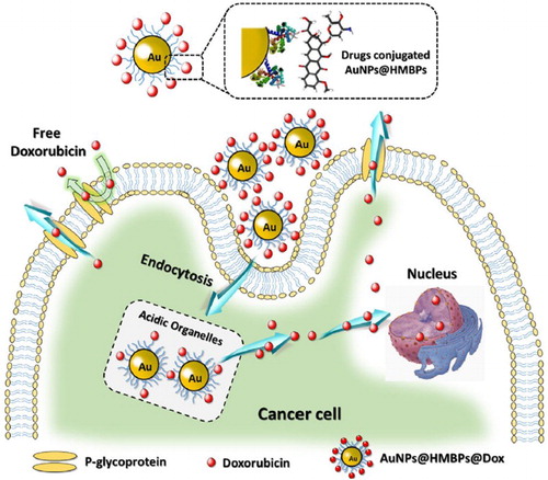
Figure 7. Biosynthesis of AuNPs by employing heavy metal binding proteins (HMBPs) in recombinant E. coli and subsequent conjugation of doxorubicin. Doxorubicin-loaded AuNPs complexes can be easily fragmented to release doxorubicin from AuNPs to defuse through the cancer cell (Adapted with permission from Seo et al. (Citation124)).

Figure 7. Biosynthesis of AuNPs by employing heavy metal binding proteins (HMBPs) in recombinant E. coli and subsequent conjugation of doxorubicin. Doxorubicin-loaded AuNPs complexes can be easily fragmented to release doxorubicin from AuNPs to defuse through the cancer cell (Adapted with permission from Seo et al. (Citation124)).

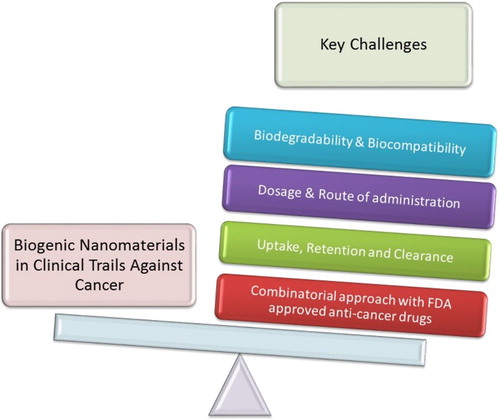
Figure 8. Major challenges for scientists before biosynthesized nanomaterials enter clinical trials for cancer theranostics.

Figure 8. Major challenges for scientists before biosynthesized nanomaterials enter clinical trials for cancer theranostics.