Figures & data
Figure 1. Change in color for leaf extract containing silver before and after the preparation of AgNPs.
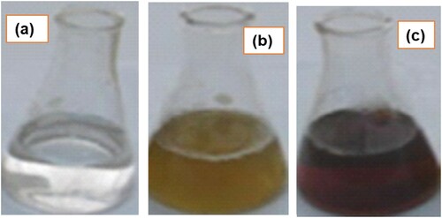
Figure 2. UV–Vis spectra analysis: Plasmon resonance of AgNP05 reduced by D. carota at 437 nm and plasmon resonance of AgNP01 reduced by D. Carota at 429 nm.
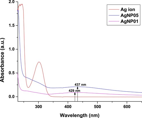
Figure 3. Comparative FTIR spectra of leaf extract, AgNP05 and AgNP01 obtained from D. carota L. (DCLE).
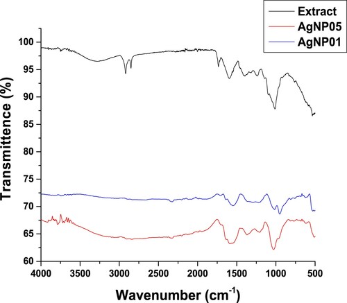
Figure 4. X-ray diffraction patterns of synthesized (a) AgNP05 and (b) AgNP01 from D. carota leaf extract (DCLE).
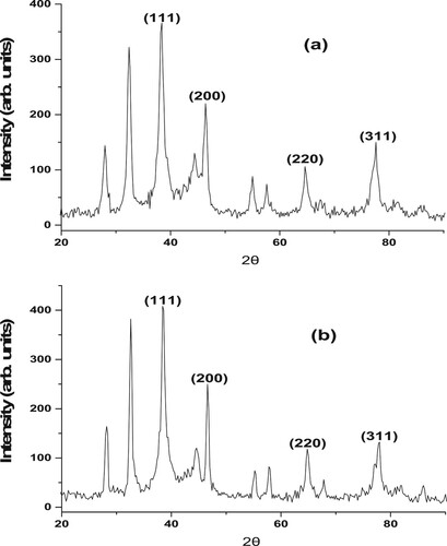
Figure 5. TEM image of the synthesized (a) AgNP05 and (b) AgNP01 from D. carota leaf extract (DCLE).
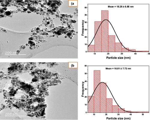
Figure 6. Mechanism of reaction for the action of silver nanoparticles (AgNP05 and AgNP01) as an antioxidant.
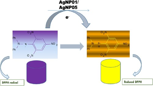
Table 1. Antioxidant activity of D carota L. leaf extract (DCLE) and silver nanoparticles against DPPH radical.
Figure 7. DPPH free radical scavenging assays of D carota L. leaf extract (DCLE) and silver nanoparticles (AgNP05 and AgNP01) (n = 3); μg/ml = microgram per millilitres.
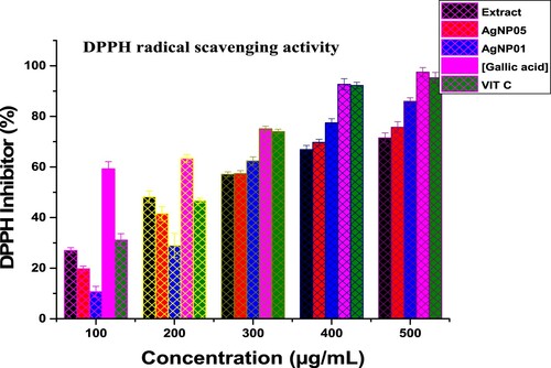
Figure 8. Antibacterial activities of D carota L. leaf extract (DCLE) and AgNPs (a), Antifungal activities of D carota L. leaf extract (DCLE) and AgNPs (b).
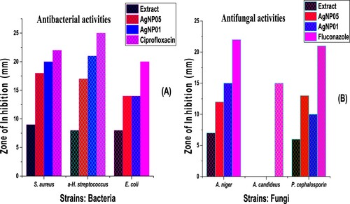
Table 2. Antimicrobial potential of D carota L. leaf extract (DCLE) and silver nanoparticles against bacteria and fungi pathogens.
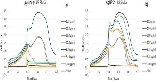
Table 3. The ED50 (μg/ml) of silver nanomaterials (AgNP05 and AgNP01) against brain glioblastoma cells, U87MG.
Figure 9. xCELLigence real-time toxicity for silver nanomaterials AgNP05 (a) and AgNP01 (b) on the U87MG cell line, at concentrations (100, 50.0, 25.0, 12.5, 6.25, 3.125 and 0 μg/ml).