Figures & data
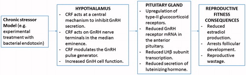
Figure 1. Diagram of the hypothalamic–pituitary–adrenal axis (HPA-axis) and hypothalamic–pituitary–gonadal axis (HPG-axis) loop in the female animal, detailing feedback and interactions between the hormones and structures involved in both pathways. Oestrogen and progesterone confer positive/negative feedback onto the hypothalamus to regulate the HPG-axis. Cortisol confers negative feedback onto the hypothalamus to regulate the HPA-axis. Cortisol also acts at the level of the anterior pituitary (P = pituitary) and reproductive organs to impede reproductive function. Hypothalamic kisspeptin (KNDy cells) are believed to modulate the synthesis of gonadotropin releasing hormone (GnRH) within the hypothalamus. Gonadotrophin inhibitory hormone (GnIH) neurons are assumed to act both directly on GnRH neurons within the hypothalamus and project to the median eminence to mediate pituitary function via G-protein coupled receptors on gonadotroph cells.
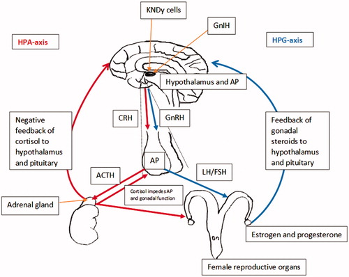
Figure 2. Diagram summarising the main neuroendocrine changes occurring in the animal (sheep model) due to direct exposure to chronic stressor, and the culminating effects on reproductive fitness of the animal (this summary diagram is based on the published literature).