985
Views
4
CrossRef citations to date
0
Altmetric
Research Papers

Identification of novel putative-binding proteins for cellular prion protein and a specific interaction with the STIP1 homology and U-Box-containing protein 1

, , , , , , , & show all
Pages 355-366 | Received 09 Mar 2015, Accepted 14 Jul 2015, Published online: 28 Aug 2015

Figures & data

TABLE 1. Gene identities obtained from yeast two-hybrid screening

Figure 1. PrPC interacts with 10 different ligands in cross-mating assay. Bait strain expressing PrPC and the Lac-Z reporter gene were mated with target strains expressing the putative PrPC interactors. X-gal was used to score positive interactions (blue color development). pBait and pTarget were used as a positive control for interaction, since they express known interaction partners. The empty bait vector (pGilda), which expresses only Lex A domain, was used as negative control.
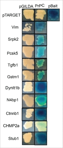
Figure 1. PrPC interacts with 10 different ligands in cross-mating assay. Bait strain expressing PrPC and the Lac-Z reporter gene were mated with target strains expressing the putative PrPC interactors. X-gal was used to score positive interactions (blue color development). pBait and pTarget were used as a positive control for interaction, since they express known interaction partners. The empty bait vector (pGilda), which expresses only Lex A domain, was used as negative control.

TABLE 2. Ten putative PrPC ligands

Figure 2. Stub1 is expressed in both olfactory epithelium (OE) and brain. (a) Protein extracts prepared from mouse brain and OE were analyzed by western blotting with anti-Stub1 (upper panel) antibodies. Anti-β-actin was used as loading control (lower panel). (b) Protein relative values were quantified using ImageJ Software® and normalized to β-actin. The results are representative of 4 independent experiments. Statistical analysis was conducted with GraphPad Prism (values were expressed as mean ±SE. Student's t test was used for statistical analysis and P<0.05 was considered significant).
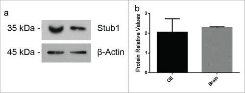
Figure 2. Stub1 is expressed in both olfactory epithelium (OE) and brain. (a) Protein extracts prepared from mouse brain and OE were analyzed by western blotting with anti-Stub1 (upper panel) antibodies. Anti-β-actin was used as loading control (lower panel). (b) Protein relative values were quantified using ImageJ Software® and normalized to β-actin. The results are representative of 4 independent experiments. Statistical analysis was conducted with GraphPad Prism (values were expressed as mean ±SE. Student's t test was used for statistical analysis and P<0.05 was considered significant).

Figure 3. PrPC interacts with Stub1. (a) A pull-down assay was performed using mouse brain extract incubated with His6-PrPC bound to Ni-NTA-agarose beads (+) or Ni-NTA-agarose alone (−). Pulled-down proteins were analyzed by western blotting (WB) employing anti-Stub1 antibody (upper panel). The same membranes subjected to pull-down assay were re-probed with anti-his-tag antibody to attest that recombinant PrPC was recovered from the beads (lower panel). HEK293T cells protein extracts overexpressing GFP-PrPC and Myc-Stub1 (b) or olfactory epithelium (OE) extracts (c) were immunoprecipitated with anti-PrPC or pre-immune serum (PI). Co-precipitated proteins were analyzed using an anti-Stub1 antibody (upper panels). The same membranes were re-probed with anti-PrPC (lower panels) to confirm that PrPC was precipitated during IP-reaction.
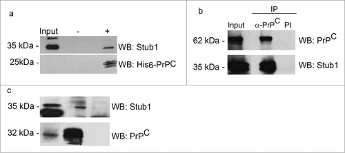
Figure 3. PrPC interacts with Stub1. (a) A pull-down assay was performed using mouse brain extract incubated with His6-PrPC bound to Ni-NTA-agarose beads (+) or Ni-NTA-agarose alone (−). Pulled-down proteins were analyzed by western blotting (WB) employing anti-Stub1 antibody (upper panel). The same membranes subjected to pull-down assay were re-probed with anti-his-tag antibody to attest that recombinant PrPC was recovered from the beads (lower panel). HEK293T cells protein extracts overexpressing GFP-PrPC and Myc-Stub1 (b) or olfactory epithelium (OE) extracts (c) were immunoprecipitated with anti-PrPC or pre-immune serum (PI). Co-precipitated proteins were analyzed using an anti-Stub1 antibody (upper panels). The same membranes were re-probed with anti-PrPC (lower panels) to confirm that PrPC was precipitated during IP-reaction.

Reprints and Corporate Permissions

Please note: Selecting permissions does not provide access to the full text of the article, please see our help page How do I view content?

To request a reprint or corporate permissions for this article, please click on the relevant link below:

Academic Permissions

Please note: Selecting permissions does not provide access to the full text of the article, please see our help page How do I view content?

Obtain permissions instantly via Rightslink by clicking on the button below:

If you are unable to obtain permissions via Rightslink, please complete and submit this Permissions form. For more information, please visit our Permissions help page.