Figures & data
TABLE 1. Summary of the 3 broad categories of prion diseases and the defining characteristics of each
FIGURE 1. PrPse propagation in the CNS. Schematic representation of the propagation of PrPsc in neuronal cells of the CNS. PrPSc acquired by infection, mutation or spontaneous conversion of cellular PrP (PrPC) combines with PrPc thereby converting it to PrPC is recruited and converted ti PrPSc. As PrPSc levels increase PrPC recruitment and conversion become more efficient, leading to an accumulation of PrPSc in neuronal cells. PrPSc accumulation causes cell dysfunction followed by death.
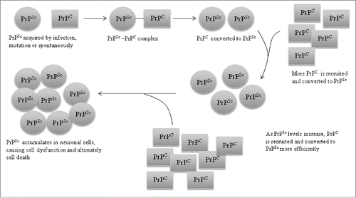
FIGURE 2. Comparison of protein structure of PrPC and PRPSc. Shown is a schematic representation of the secondary structure of PrPC in comparison to PrPSc as inferred from a recombinant fragment of Syrian hamster PrP comprising amino cid residues 90-231.A,B and C indicate α-helices.
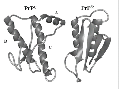
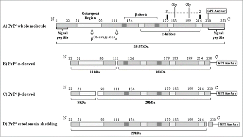
FIGURE 3. Schematic representation of the protein structure and the proteolytic processing of the PrPC in human cells. (A) Whole PrPC moleule. Post-translational modification is initiated by the removal of N-terminal and C-terminal signal p eptides. (B) Normal constitutive cleavage occurring in brain and culture cells. (C) β-cleavage mediated by reactive oxygen species (ROS). (D) Ecotodomain Shecking of the PrPC where nearly whole PrPc molecule is released from the cellular membrane.