
Figures & data
FIGURE 1. Comparison of PrPSc in brains digested with various PK concentrations. PrPSc was detected with anti-PrP mAb T2. The PK concentrations are shown on the top of each lane. The % PrPSc values are indicated at the bottom of each lane. Molecular marker sizes are indicated on the left.
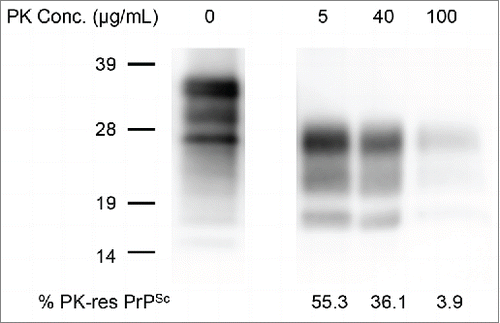
Table 1. Comparison of survival days between mice inoculated with crude homogenate and the PK-digested mBSE homogenates
FIGURE 2. Pathological changes in the brain occurring during mBSE disease progression. Groups of mice were sacrificed at the defined time points indicated on the left side of the panels. Fixed brain sections from each culling time were subjected to hematoxylin and eosin staining (B, D, F, H, J, and L) together with immunohistochemistry for PrPSc (A, C, E, G, I, and K). PrPSc immunoreactivities were detected with the anti-PrP monoclonal antibody SAF84. Micrographs show the dorsal medulla (A, B, E, F, I, and J) and cerebral cortex (C, D, G, H, K, and L).
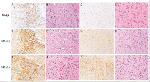
FIGURE 3. Detection of PrPSc in the brains of mice by western blot. PrPSc was detected with anti-PrP mAb T2. The defined time points (days post-inoculation, dpi) are indicated on the top of the blot. Mo and Nl indicate mouse scrapie ObihiroCitation39 and the uninfected mouse brain, respectively. The relative protein load in each lane is indicated on the bottom; 1x indicates 0.25 mg brain equivalent. Molecular markers are indicated on the left side of the blot.
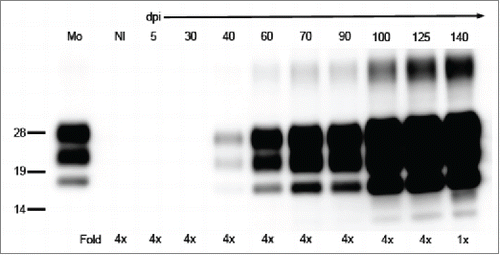
FIGURE 4. Kinetics of mBSE prion replication and PrPSc accumulation in the mouse brain during disease progression. Black circles and blue squares show the infectious units and PrPSc accumulation in the mouse brains. The infectious dose of mBSE prion at each time point was estimated by the incubation period bioassay method. ELISA was used for measuring the amount of PrPSc at the same time points, and the relative values (%) are indicated against the amount of PrPSc in the brain at the terminal stage. The kinetics of mBSE prion replication and PrPSc accumulation are shown as a semi-logarithmic plot. An exponential correlation was observed between the incubation time and mBSE prion infectivity from 5 to 100 dpi (y = 33557* e^(0.082279x) R2 = 0.94593, where y is the infectious unit and x is the survival days). A similar correlation was observed between the incubation time and PrPSc accumulation from 40 to 140 dpi (y = 0.024371* e^(0.056019x) R2 = 0.90149, where y is the relative value (%) compared to the PrPSc values of 140-dpi brains and x is the survival days).
FIGURE 5. Comparison of PrPSc and prion infectivity between the crude homogenate and 20K-sup. (A) Comparison of PrPSc amounts between the crude homogenate and 20K-sup. The relative protein load in each lane is indicated on the top. Nl indicates the uninfected mouse brain. PrP was detected with anti-PrP mAb SAF84; 1x indicates 0.25 mg brain equivalent. Molecular markers are indicated on the left side of the blot. (B) The relationship between incubation time (mean days ± standard error) and infectious units (log LD50/gram of the brain) in mice inoculated with serial dilutions of the crude homogenate (black circles and solid line; y = 16.642 – 0.061312x, where y is log LD50 and x is the incubation period) and the 20K-sup (blue squares and dotted line y = 17.474 – 0.060916x).
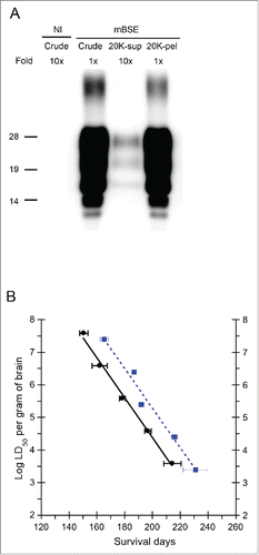
Table 2. Comparison of mBSE prion infectious units between the crude homogenate and 20K-sup by an endpoint titration assay