1,347
Views
15
CrossRef citations to date
0
Altmetric
Extra Views

Functions of the cellular prion protein, the end of Moore's law, and Ockham's razor theory

&
Pages 25-40 | Received 23 Oct 2015, Accepted 24 Nov 2015, Published online: 04 Apr 2016

Figures & data

FIGURE 1. Evolution of Moore's law from 1971 to 2011. Source: https://commons.wikimedia.org/wiki/User:Wgsimon through Creative Commons Attribution-ShareAlike License (https://creativecommons.org/licenses/by-sa/3.0/).

FIGURE 1. Evolution of Moore's law from 1971 to 2011. Source: https://commons.wikimedia.org/wiki/User:Wgsimon through Creative Commons Attribution-ShareAlike License (https://creativecommons.org/licenses/by-sa/3.0/).

FIGURE 2. (A) Domain organization of PrPC (mouse sequence). (B) Examples of some derived truncated forms used in in vitro and in vivo studies. The effects of their transfection in cells are indicated from (++) strong effect to negligible (−) effects in apoptosis. The examples of truncated PrPC forms are summarized from.Citation68,94,106,107 Results obtained in several studies reinforced data obtained by D.R. Brown's Lab in 2003.Citation108(C) Epitope mapping of some antibodies used in cytotoxicity studies. The name and recognized PrPC region is indicated in each case. Green antibodies indicate that their use is non-cytotoxic in contrast to red antibodies.
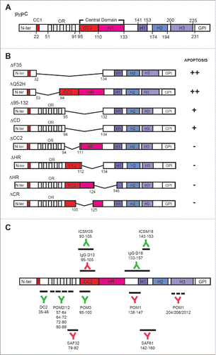
FIGURE 2. (A) Domain organization of PrPC (mouse sequence). (B) Examples of some derived truncated forms used in in vitro and in vivo studies. The effects of their transfection in cells are indicated from (++) strong effect to negligible (−) effects in apoptosis. The examples of truncated PrPC forms are summarized from.Citation68,94,106,107 Results obtained in several studies reinforced data obtained by D.R. Brown's Lab in 2003.Citation108(C) Epitope mapping of some antibodies used in cytotoxicity studies. The name and recognized PrPC region is indicated in each case. Green antibodies indicate that their use is non-cytotoxic in contrast to red antibodies.

FIGURE 3. Scheme illustrating the effect of the expression of particular truncated forms of PrPC (B-E), treatment with GD-directed antibodies (F), peptides recognizing the CD region (G), aggregating antibodies recognizing GD and OR regions (H), and pathogenic prion protein (I). Absence of the OR in B and C leads to increased apoptosis. In contrast, PrPC lacking the CD but more relevantly lacking both the GD and the CD induced increased neurotoxicity. In contrast, aggregating antibodies (H), GD-directed antibodies (F), peptides (G), and the pathogenic prion (I) lead to profound changes in the 3D organization of PrPC in the membrane, which triggers the approach of the N-terminal region to the plasma membrane (red curved arrow) leading to increased ROS production and cell death as observed in PrPC constructs with artificial tethering of the N-terminal to the membrane. In these conditions, PrPC recycling is very low and their homeostatic function is lost.
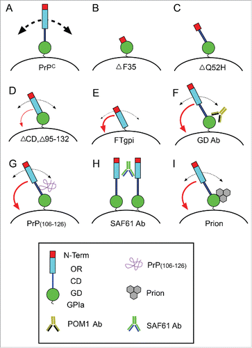
FIGURE 3. Scheme illustrating the effect of the expression of particular truncated forms of PrPC (B-E), treatment with GD-directed antibodies (F), peptides recognizing the CD region (G), aggregating antibodies recognizing GD and OR regions (H), and pathogenic prion protein (I). Absence of the OR in B and C leads to increased apoptosis. In contrast, PrPC lacking the CD but more relevantly lacking both the GD and the CD induced increased neurotoxicity. In contrast, aggregating antibodies (H), GD-directed antibodies (F), peptides (G), and the pathogenic prion (I) lead to profound changes in the 3D organization of PrPC in the membrane, which triggers the approach of the N-terminal region to the plasma membrane (red curved arrow) leading to increased ROS production and cell death as observed in PrPC constructs with artificial tethering of the N-terminal to the membrane. In these conditions, PrPC recycling is very low and their homeostatic function is lost.