2,374
Views
24
CrossRef citations to date
0
Altmetric
Extra Views

Prions on the run: How extracellular vesicles serve as delivery vehicles for self-templating protein aggregates

, , &
Pages 98-112 | Received 16 Feb 2017, Accepted 07 Mar 2017, Published online: 12 Apr 2017

Figures & data

FIGURE 1. Cell culture assays to study prion infection by exosomes. (A) Cell culture assays to study exosome-mediated TSE prion infection. Published exosome-mediated TSE infection assays are time consuming und rely on the detection of newly formed, proteinase K (PK) resistant PrPSc. Naive cells permissive to infection with the respective TSE prion strain are exposed to exosomal preparations isolated from prion-infected cells for 4–5 days, followed by several weeks of culture. Read-out is PK-resistant PrPSc detected by cell blot or western blot.Citation42,47-49,51 (B) Quantitative imaging of exosome-mediated NM aggregate induction. Recipient NM-GFPsol cells are seeded on a 384 well plate for 1 hour. Exosomes isolated from conditioned medium of donor cells are added to the wells. Life or fixed cells are subjected to automated high throughput confocal microscopy. Read-out is induction of NM-GFP aggregates in recipient cells. Life imaging analysis demonstrates the appearance of NM-GFP aggregates as soon as 3 hours post exosome addition. The arrowhead marks cells with exosome-induced NM-GFP aggregates. The assay can also reveal bidirectional inheritance of NM aggregates by daughter cells, a characteristic of TSE prions replicating in cellular models.Citation6
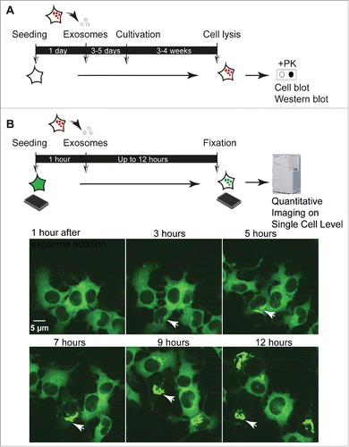
FIGURE 1. Cell culture assays to study prion infection by exosomes. (A) Cell culture assays to study exosome-mediated TSE prion infection. Published exosome-mediated TSE infection assays are time consuming und rely on the detection of newly formed, proteinase K (PK) resistant PrPSc. Naive cells permissive to infection with the respective TSE prion strain are exposed to exosomal preparations isolated from prion-infected cells for 4–5 days, followed by several weeks of culture. Read-out is PK-resistant PrPSc detected by cell blot or western blot.Citation42,47-49,51 (B) Quantitative imaging of exosome-mediated NM aggregate induction. Recipient NM-GFPsol cells are seeded on a 384 well plate for 1 hour. Exosomes isolated from conditioned medium of donor cells are added to the wells. Life or fixed cells are subjected to automated high throughput confocal microscopy. Read-out is induction of NM-GFP aggregates in recipient cells. Life imaging analysis demonstrates the appearance of NM-GFP aggregates as soon as 3 hours post exosome addition. The arrowhead marks cells with exosome-induced NM-GFP aggregates. The assay can also reveal bidirectional inheritance of NM aggregates by daughter cells, a characteristic of TSE prions replicating in cellular models.Citation6

FIGURE 2. Human HEK cells secrete both soluble and aggregated Sup35 NM in association with exosomes. HEK cells expressing HA-epitope tagged NM (NM-HA) before (A) and after (B) NM aggregate induction by recombinant NM fibrils. NM-HAsol: soluble NM-HA. NM-HAagg: aggregated NM-HA. NM-HA was stained with anti-HA antibody (red) and nuclei were counterstained with Hoechst (blue). Maximum intensity projections were generated from Z-stacks. (C) Western blot analysis of exosomes from HEK NM-HAsol, NM-HAagg and N2a NM-HAagg s2E cell clones for exosomal marker Alix and NM-HA. Exosomes were isolated according to a previously described method.Citation72 (D) Exosome numbers released from HEK NM-HAsol and NM-HAagg were determined using ZetaView PMX 110-SZ-488 Nano Particle Tracking Analyzer with the same measurement setting. Results shown are means ± SD (n = 3; *** p < 0.001; unpaired student t test). (E) Exosome numbers released from HEK NM-HAagg, N2a NM-HAagg clone s2E (selected for high aggregate inducing activity in recipient cells) and N2a NM-HAagg clone 1C. Results shown are means ± SD (n = 3; *** p<0.001; one-way ANOVA). (F) Glutaraldehyde cross-linking of proteins in cell lysates or exosomes from HEK NM-HAsol, HEK NM-HAagg and N2a NM-HAagg clones s2E or 1C to determine the oligomerization state of NM-HA. Cross-linking was done as described previously.Citation72
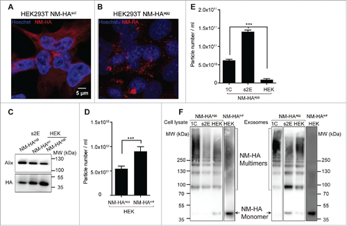
FIGURE 2. Human HEK cells secrete both soluble and aggregated Sup35 NM in association with exosomes. HEK cells expressing HA-epitope tagged NM (NM-HA) before (A) and after (B) NM aggregate induction by recombinant NM fibrils. NM-HAsol: soluble NM-HA. NM-HAagg: aggregated NM-HA. NM-HA was stained with anti-HA antibody (red) and nuclei were counterstained with Hoechst (blue). Maximum intensity projections were generated from Z-stacks. (C) Western blot analysis of exosomes from HEK NM-HAsol, NM-HAagg and N2a NM-HAagg s2E cell clones for exosomal marker Alix and NM-HA. Exosomes were isolated according to a previously described method.Citation72 (D) Exosome numbers released from HEK NM-HAsol and NM-HAagg were determined using ZetaView PMX 110-SZ-488 Nano Particle Tracking Analyzer with the same measurement setting. Results shown are means ± SD (n = 3; *** p < 0.001; unpaired student t test). (E) Exosome numbers released from HEK NM-HAagg, N2a NM-HAagg clone s2E (selected for high aggregate inducing activity in recipient cells) and N2a NM-HAagg clone 1C. Results shown are means ± SD (n = 3; *** p<0.001; one-way ANOVA). (F) Glutaraldehyde cross-linking of proteins in cell lysates or exosomes from HEK NM-HAsol, HEK NM-HAagg and N2a NM-HAagg clones s2E or 1C to determine the oligomerization state of NM-HA. Cross-linking was done as described previously.Citation72

FIGURE 3. Factors influencing seeding activity of protein aggregates incorporated into exosomes. Exosome-mediated secretion of NM-HA by donor cells and subsequent uptake and seeding of NM-GFP prions in recipient cells. Infectivity of NM-HA bearing exosomes is likely determined by the following parameters: (1) Enhanced secretion of exosomes, (2, 3) Selective sorting of low-order oligomers, (4, 5) Specific exosome-target cell interaction (ligand-receptor recognition). This could include cell specific ligand-receptor interactions or differences in the intracellular fate of endocytosed exosomes. After internalization, the NM-HA aggregates contained in exosomes are released and induce new aggregate formation in N2a NM-GFP cells. The mechanism of NM-HA release into the cytosol is so far unknown.
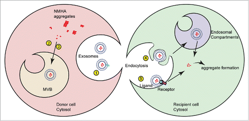
FIGURE 3. Factors influencing seeding activity of protein aggregates incorporated into exosomes. Exosome-mediated secretion of NM-HA by donor cells and subsequent uptake and seeding of NM-GFP prions in recipient cells. Infectivity of NM-HA bearing exosomes is likely determined by the following parameters: (1) Enhanced secretion of exosomes, (2, 3) Selective sorting of low-order oligomers, (4, 5) Specific exosome-target cell interaction (ligand-receptor recognition). This could include cell specific ligand-receptor interactions or differences in the intracellular fate of endocytosed exosomes. After internalization, the NM-HA aggregates contained in exosomes are released and induce new aggregate formation in N2a NM-GFP cells. The mechanism of NM-HA release into the cytosol is so far unknown.