
Figures & data
FIGURE 1. MRI and FDG-PET. In upper row, diffusion-weighted imaging with no alterations. In middle row, fluid attenuated inversion recovery (FLAIR) sequence showing global atrophy. In lower row, FDG-PET imaging showed a left frontal-parietal, bilateral thalamus and cerebellar hypometabolism.
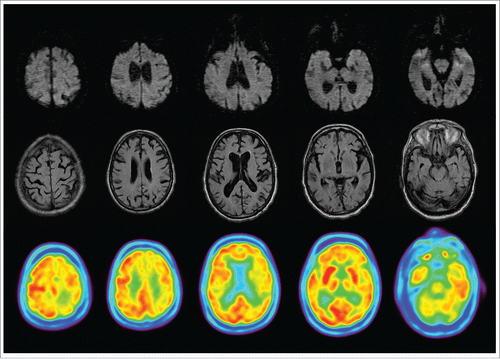
FIGURE 2. 18F-florbetaben PET imaging. Uptake tracer in white matter is generally higher than in gray matter. In left parietal and right superior temporal lobes, tracer uptake is similar in white and gray matter.
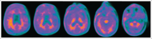
FIGURE 3. Frontal cortex showing no deposition of β-amyloid.
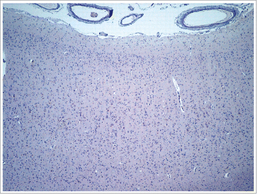
FIGURE 4. Pathological prion protein expression in several brain regions. Immunoreactivity of pathological prion protein in 6 regions (inferior temporal, caudate, anterior cingulate, calcarine, supplementary motor area, and precuneus) is shown. Each region is observed at 1.5x and 20x magnification. Different levels of expression (low, medium, and high) may be observed.
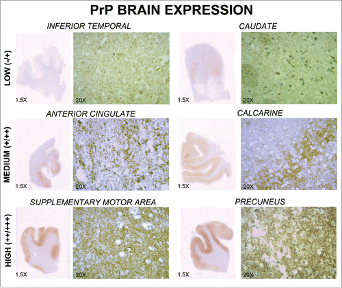
TABLE 1. Studies of prion diseases using amyloid PET tracers.