Figures & data
TABLE 1. Patient characteristics, codon 129 polymorphism, presence of periodic sharp wave complexes on electroencephalogram and CSF 14-3-3 protein, associated clinical signs/syndromes, time between symptom onset and PET, and lateralization of PET hypometabolism.
TABLE 2. FDG-PET data for the entire (global) CJD group and the different clinical CJD subgroups.
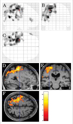
FIGURE 1. SPM showing decreased FDG-PET metabolism (using a stringent level of significance of p<0.001 and an extent threshold of 100 voxels) in the left lateral frontal cortex and the left mesial parietal cortex, and to a lesser degree the right mesial parietal cortex left lateral posterior temporal cortex in CJD patients in comparison to controls.
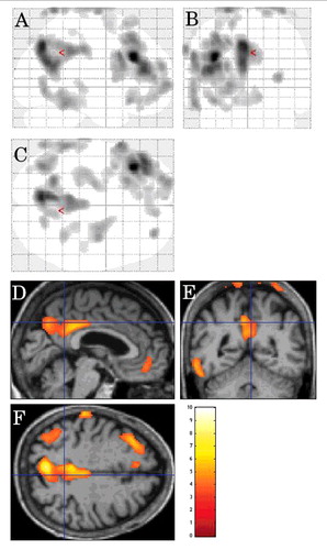
FIGURE 2. Inter-CJD SPM analysis, using a stringent level of significance of p<0.001 and an extent threshold of 100 voxels, in patients with cerebellar ataxia, showing FDG-PET hypometabolism in the pons, the bilateral (left-predominant) middle cerebellar peduncles, the right frontal subcortical lobe, and the left mesial temporal cortex.
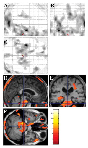
FIGURE 3. Inter-CJD SPM analysis showing decreased metabolism (using a stringent level of significance of p<0.001 and an extent threshold of 100 voxels) in the bilateral (right-predomiant) occipital cortex and the right frontal subcortical lobe in CJD patients with visual signs.
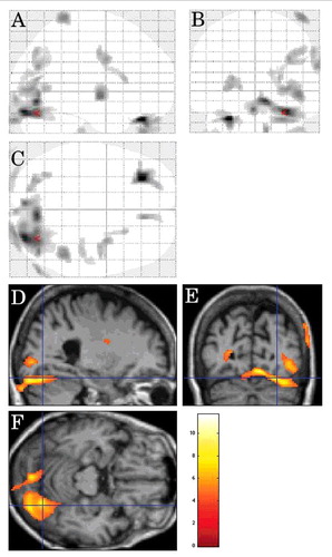
FIGURE 4. Inter-CJD SPM analysis (using a stringent level of significance of p<0.001 and an extent threshold of 100 voxels), in CJD patients with myoclonus, showing hypometabolism in the left lateral parietal cortex.
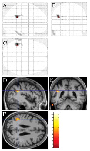
FIGURE 5. Inter-CJD SPM analysis, in the CJD patients with limb dystonia, showing left lateral parietal cortex and left lateral prerolandic frontal cortex hypometabolism (using a stringent level of significance of p<0.001 and an extent threshold of 100 voxels).
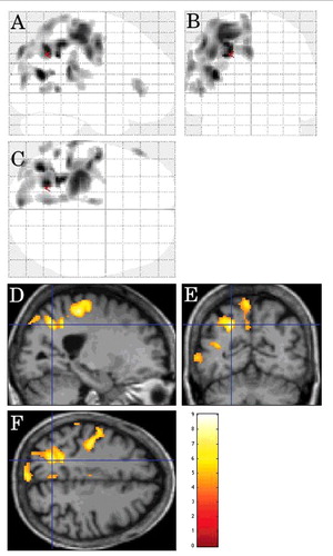
FIGURE 6. Inter-CJD SPM analysis showing hypometabolism in the left lateral prerolandic frontal cortex, the left -lateral more than mesial- parietal cortex, and the left lateral posterior temporal cortex in patients with corticobasal syndrome.