Figures & data
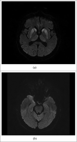
Figure 1. (A). MRI DWI: abnormal high signal intensity in both caudate nuclei, putamina and thalami, particularly prominent in heads of caudate nuclei and anterior parts of putamina. (B). MRI DWI: slight changes in cortex of right temporal lobe and colliculi inferior.
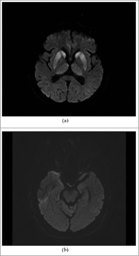
Figure 2. (A). MRI DWI: abnormal high signal intensity in both caudate nuclei, putamina and thalami, particularly prominent in heads of caudate nuclei and anterior parts of putamina (the same changes as seen on Fig. A). (B). MRI DWI: abnormal high signal intensity in cortex of anterior pole of the right temporal lobe and slight, linear hyperintensity in cortex of the left temporal lobe.