Figures & data
Figure 1. Chronologic timeline of the clinical manifestations, the results of examinations, main therapeutics and diagnosis of this P102 GSS case.
Figure 2. Diffusion weighted imaging (DWI) MRI. High intense signal changes in bilateral frontal, parietal, temporal and occipital cortices.
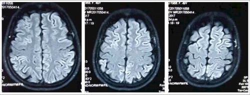
Figure 3. Western blot for CSF 14-3-3 and PRNP gene sequencing. A. Western blot. The tested CSF samples were separated in 12% SDS-PAGE and immunobloted with 14-3-3 specific polyclonal antibody. Positive Ctrl: 10% goat brain homogenate; M: molecular weight; sCJD: CSF sample of a sCJD case; P102L GSS: CSF sample of the case in this study. B. Graphic presentation of the sequencing analysis of PRNP. A missense mutation at codon 102 (CCG to CTG) causing the substitution of Pro (P) to Leu (L) (left panel), Met (M) homozygote at codon 129 (middle panel) and Glu (E) homozygote at codon 219 (right panel).
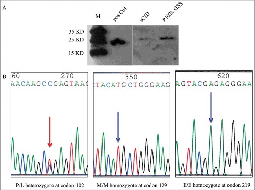
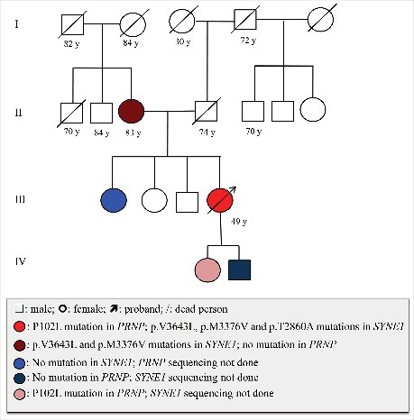
Figure 4. Medical history, P102L mutation in PRNP and the mutation in SYNE1 in the patient's family. Open square: male; open circle: female; square or circle with prolonged diagonal lines: deceased individuals; red circle with arrow: proband case with P102L mutation in PRNP and p.V3643L, p.M3376V, p.T2860A mutations in SYNE1; Dark brown circle: with p.V3643L, p.M3376V mutations in SYNE1, no mutation in PRNP; Blue circle: no mutation in SYNE1, PRNP sequencing not done; Dark blue circle: no mutation in PRNP, SYNE1 sequencing not done; Pink circle: with P102L mutation in PRNP, SYNE1 sequencing not done.