Figures & data
Figure 13. Three autophagic vacuoles in different stages of formation. The left one presents two adjacent membranes marked with black ovals; those membranes form a loop. The middle autophagic vacuole contains three electron-lucent vesicles (additional autophagic vacuoles ?); original magnification, x 33,000.
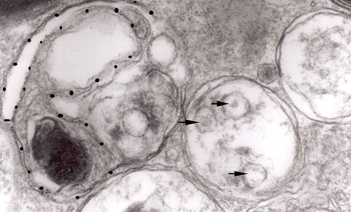
Figure 1. (a, b) An axon in an early phase of degeneration. There are numerous slender mitochondria, a multivesicular body (short arrow) and a small autophagic vacuole (arrowhead). Numerous lucent vesicles and dense-core vesicles (bent arrows) are observed. Original magnification, x 8300.
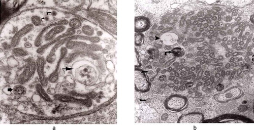
Figure 2. Degenerating axon accumulating some elongated mitochondria, ‘regular’ and ‘irregular’ MVB (arrows) and early autophagic vacuole (arrowhead pointing toward the autophagosome membrane); original magnification, x 8300.
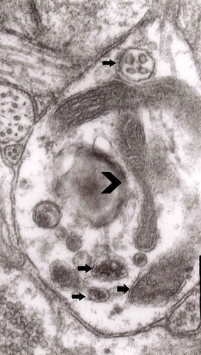
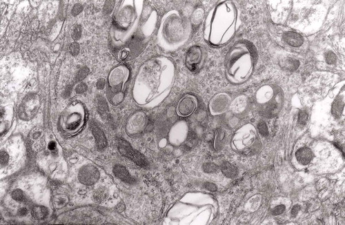
Figure 7. A fragment of a dystrophic axon containing numerous dense bodies and ‘forming and formed’ autophagic vacuoles; original magnification, 6600. This figure corresponds to Matthews Fig. 22. The details are shown in and .
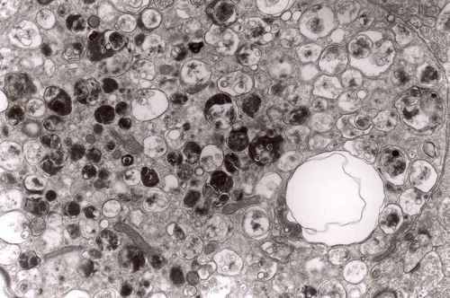
Figure 3. Dilated myelinated axons filled with a large number of MVB (arrows). This electron-micrograph is analogous to . of Matthews [Citation46]. Note that some MVB take part in a formation of autophagic vacuoles and , original magnification, x 33 000.
![Figure 3. Dilated myelinated axons filled with a large number of MVB (arrows). This electron-micrograph is analogous to Figure 16. of Matthews [Citation46]. Note that some MVB take part in a formation of autophagic vacuoles and Figure 5, original magnification, x 33 000.](/cms/asset/7a6e0060-5173-4f14-91b5-9da6037eec88/kprn_a_1595315_f0003_oc.jpg)
Figure 4. Higher magnification of part of dystrophic neurite depicted in . Note that some ‘irregular’ MVB take part in autophagic vacuole formation.
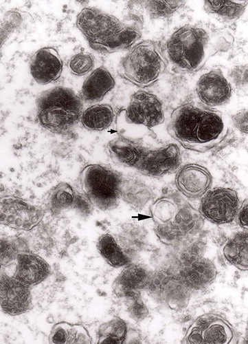
Figure 5. A large magnification of numerous ‘irregular’ MVB (arrows). This electron micrograph corresponds to . of Matthews. Note that MVB marked with double arrows presents a dark part, probably an autophagic vacuole. Original magnification, x 20 000.
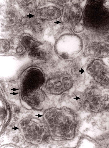
Figure 6. A fragment of a dystrophic axon containing three autophagic vacuoles with content (arrows) of ‘different stages of darkening and degradation’, original magnification, x 33,000. This figure corresponds to Matthews .
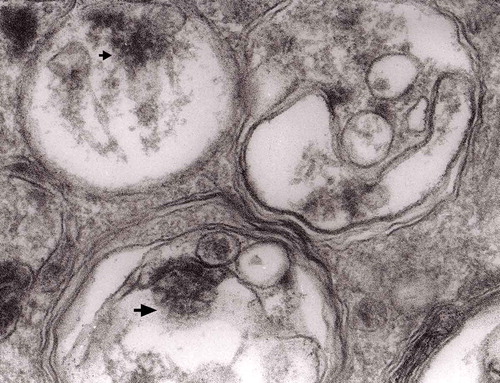
Figure 8. A fragment of . Note autophagic vacuoles (arrows) with partially electron-dense content; original magnification, x 6600.
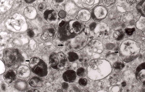
Figure 9. Another fragment of . Note two larger autophagic vacuoles (arrows) and a complex dense body. Note also, that larger vacuole of those two contains electron-dense fragment, this corresponds to partially digested content.
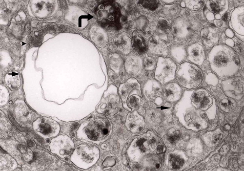
Figure 16. A typical round ‘regular’ MVB in unmyelinated nerve process; original magnification, x 16,000.
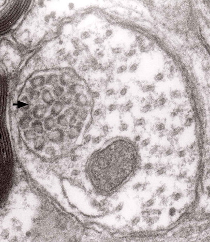
Figure 19. Accumulation of numerous MVBs of irregular sizes and partially flattened; original magnification, x 33,000.
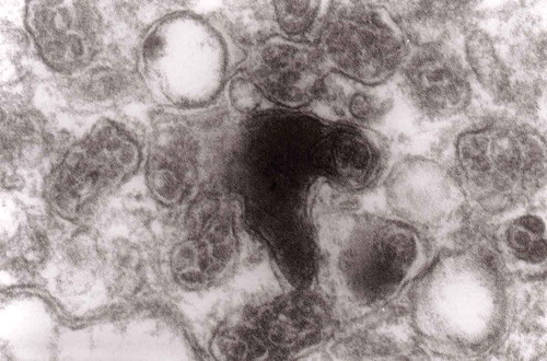
Figure 21. Another fragment of cytoplasm of the macrophage filled with numerous lysosomes and autophagic vacuoles, original magnification, x 16 000.
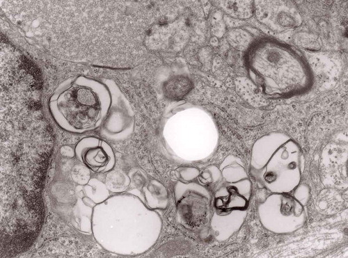
Figure 10. An enlarged fragment of dystrophic neurite showing an autophagic vacuole lined with double membrane. Original magnification, x 33,000.
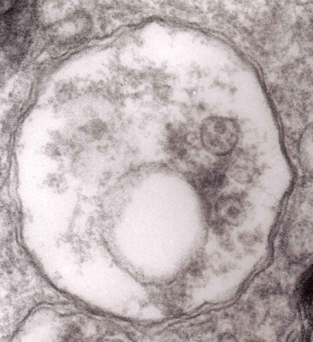
Figure 11. An enlarged fragment of dystrophic neurite showing a complex autophagic vacuole with a darkened content. Original magnification, x 33,000.
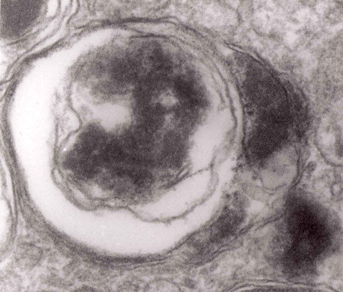
Figure 12. An enlarged fragment of dystrophic neurite showing a complex autophagic vacuole with the formed loop (two arrows) penetrating the adjacent cytoplasm. Original magnification, x 33,000.
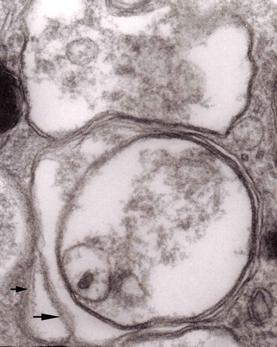
Figure 14. Several autophagic vacuoles. Note the one marked with arrows – it demonstrates elongated protrusions in a process of encircling another vacuole (bent arrow). A complex autophagic vacuole is visible in the vicinity (semicircular arrow); original magnification, x 33,000.
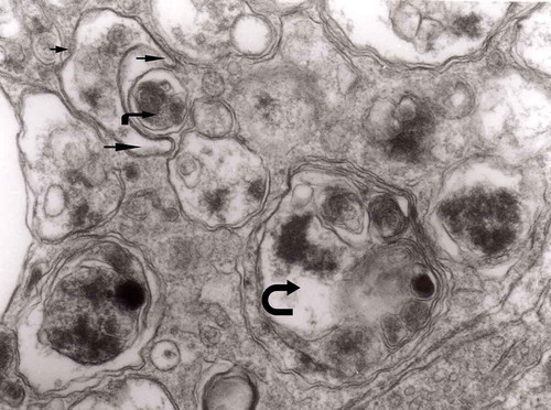
Figure 15. Accumulation of dense bodies – the similarities or even identity with autophagic vacuoles is noticeable; original magnification, x 20,000.
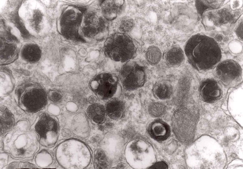
Figure 17. A dystrophic axon with numerous complex MVB (arrows). Those MVB marked with arrows contained vesicles of increased electron density while others (semicircular arrow) contain electron-lucent vesicles; original magnification, x 16,000.
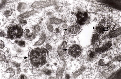
Figure 18. Enlarged fragment of the previous electron-micrograph. Note MVBs – one of normal electron density (semicircular arrow) and one with increased density (arrow); original magnification, x 16,000.
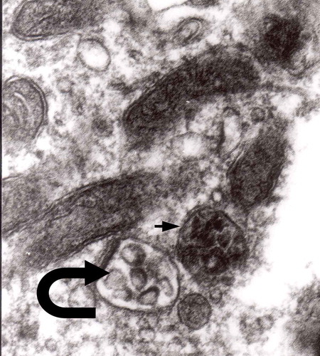
Figure 20. Cytoplasm of the macrophage filled with numerous lysosomes and autophagic vacuoles, original magnification, x 16 000.